

Musculoskeletal Clinical Solution
Musculoskeletal (MSK) Imaging is moving beyond muscles and tendons. For example, identifying the exact location of very small foreign bodies close to moving structures of the MSK anatomy is important when planning best method of removal. Another indicator of pathology in MSK studies is the presence of increased blood flow due to inflammation. Accurate detection of increased blood flow can help in monitoring the effect of therapy and in patient management.
Canon Medical’s new and improved matrix technology (iDMS) allows more control over the beam to provide a super thin slice thickness in both planes for outstanding high resolution imaging, in focus all the way from the near to far field. This new generation of matrix transducers is unique to Canon Medical and an advancement on already existing matrix technology that we have utilised for many years.
Our revolutionary SMI™ allows the visualisation of the micro-vascularity of MSK structures which can be impossible to detect with conventional Colour Doppler. SMI™ provides more clinically useful information in MSK examinations and has set a new standard in detecting micro-vascularity.
Canon Medical continues this revolution with the introduction of the new 33MHz transducer. This ultra-high frequency transducer is able to demonstrate detail in the skin layers, as well as superficial ligaments and tendons that has up until now been impossible to see.
High frequency transducers
Hand and finger ultrasound
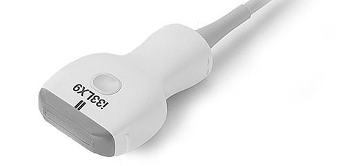
Ultra-High Frequency. Perfect For Small Parts Examinations.
33MHz Ultra-high Frequency Linear Transducer
Canon Medical’s new i33LX9 matrix linear transducer uses iDMS technology with a frequency band of up to 33MHz, which provides extremely fine detail in the near field, making it especially useful for MSK, Rheumatology and dermal imaging examinations.
Amazing Detail, Outstanding Versatility.
24MHz Ultra-high Frequency Linear Transducer
Amazing Detail, Outstanding Versatility.
24MHz Ultra-high Frequency Linear Transducer
Aplio’s i-series 24MHz transducer with extended bandwidth and iDMS technology provides excellent detail and definition in the near field, making it possible to visualise smaller structures such as nerves and small foreign bodies in MSK examinations.
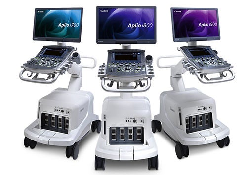
Video left: Biceps Insertion with Aplio i-series 800 system and 24MHz transducer

''The clarity and depth we can achieve with the Aplio system has transformed our approach to MSK and nerve imaging. Once challenging nerve assessments are now much more reliable and insightful.''
Ian Stewart
Chief Sonographer
The Radiology Clinic
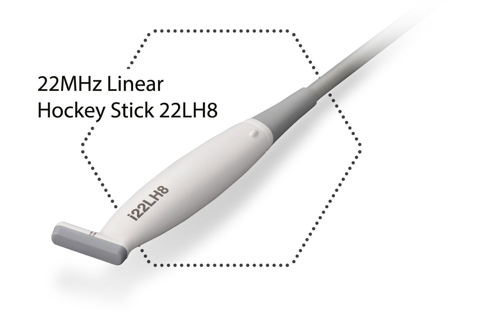
22MHZ Hockey Stick*
Ultra-high frequency, small footprint.
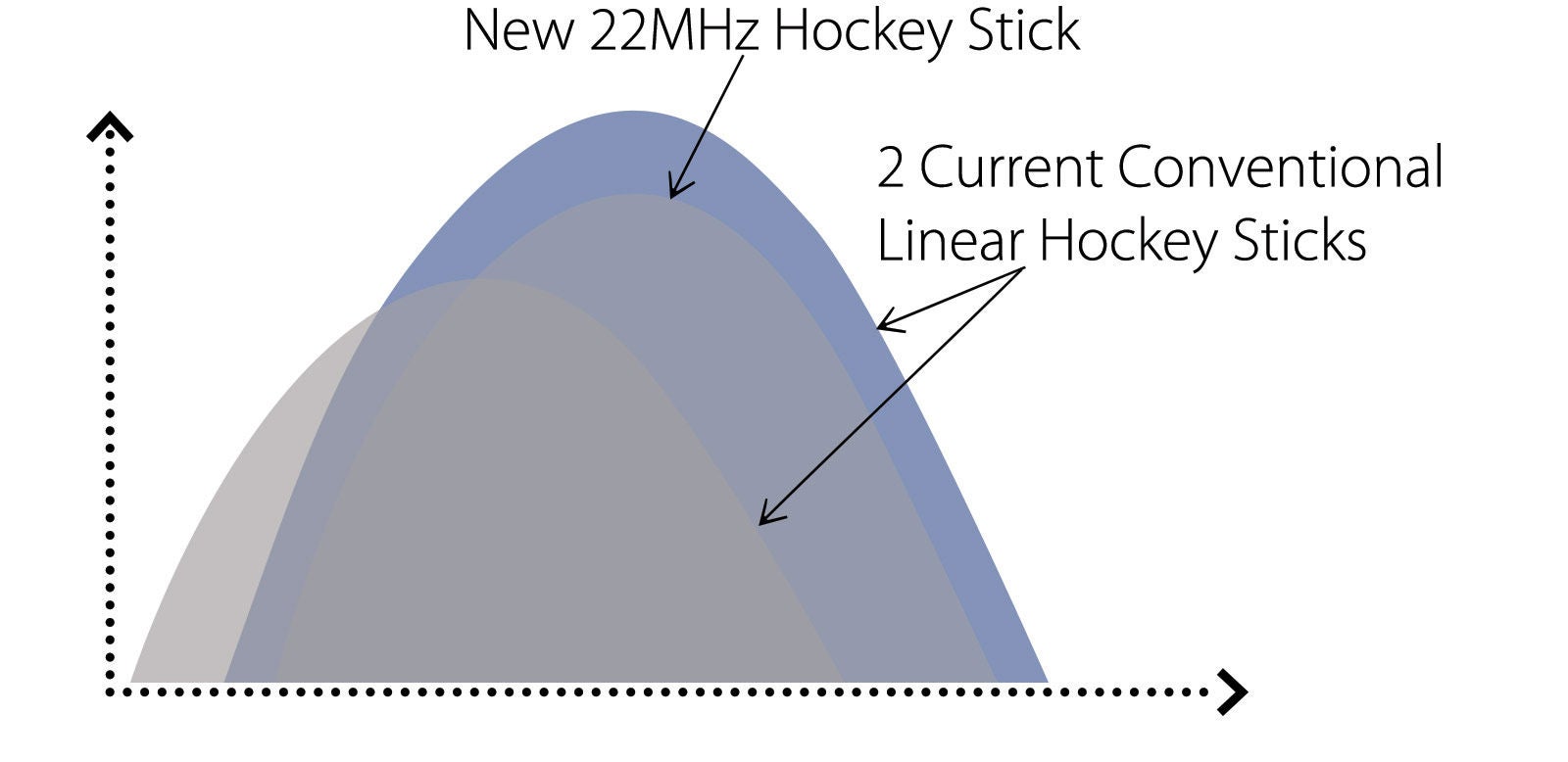
Aplio’s i-series 22MHz Hockey Stick provides the ultra-high frequency needed for the most demanding small parts examinations.
New Features:
- Very high Resolution
- Ergonomic design with small 8mm footprint and slim cable, great for examining collateral ligaments and other finger and hand pathology
- SMI-capable
Clinical Applications:
- MSK
- Small Organ
- Peripheral Vessel
- Intra-operative
- Interventional
- Compatible with the AplioTM i-series 800 system
Superb Micro-Vascular Imaging
Canon Medical Systems’ innovative Superb Micro-Vascular Imaging (SMI) technology expands the range of visible blood flow and provides visualisation of low velocity microvascular flow never before seen with ultrasound. This has proven to be valuable in the diagnosis of tendon inflammation and arthritis.
» SMI has improved dramatically to visualise the micro-vascular changes within tumours using the abdominal and linear transducers, adding more diagnostic information.«
MSK Scanning and Intervention Made Easy
» I was especially impressed by the B-mode image produced by the high quality matrix probes on Aplio i-series 800, the improved resolution has enabled us to provide high level musculoskeletal procedures which are at this stage unique to our practice – including percutaneous finger pulley release and foreign body removal, each performed with real time ultrasound guidance on this platform.«
Matthew Bondarenko, Ultrasound Team Leader,
Benson Radiology Ashford Hospital Adelaide
Healthy Sonographer Program
Surveys show 90% of sonographers work in pain and sadly, 20% suffer career ending injuries**
Watch the experts demonstrate the latest innovations in ergonomics that improve sonographer comfort in order to help reduce risk of injury.
- *Excerpt from: Sonographer Safety Workplace Conditions
Clinical images
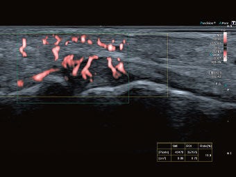
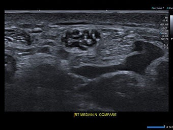
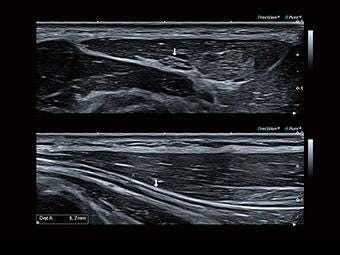
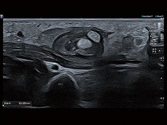
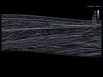
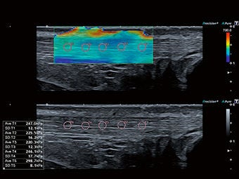
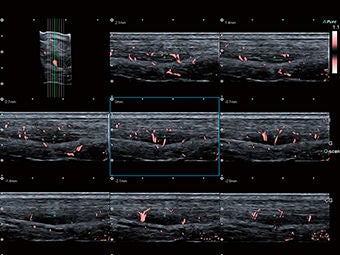
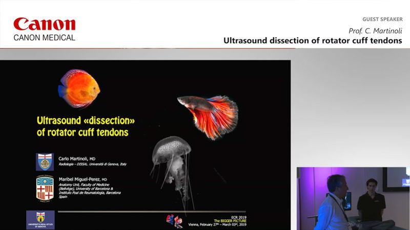
ECR2019 Workshops
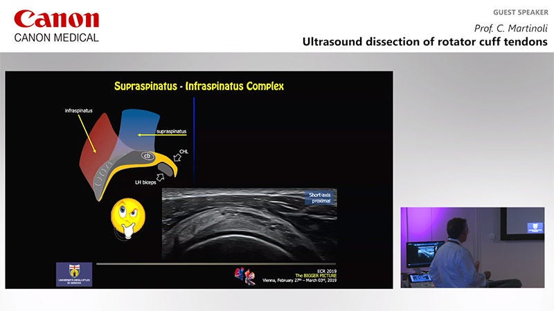
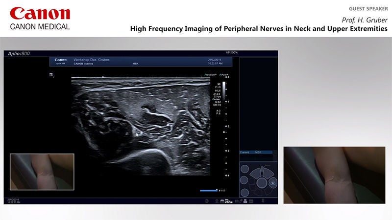
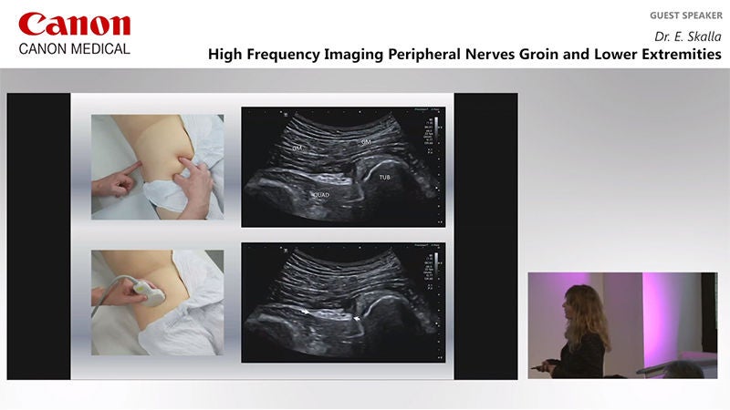
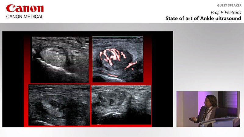
Education and Resources
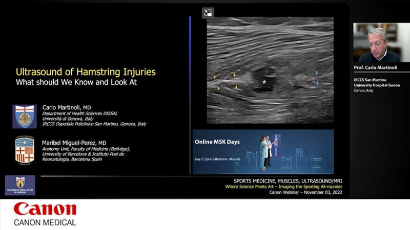
Interview report & Case study
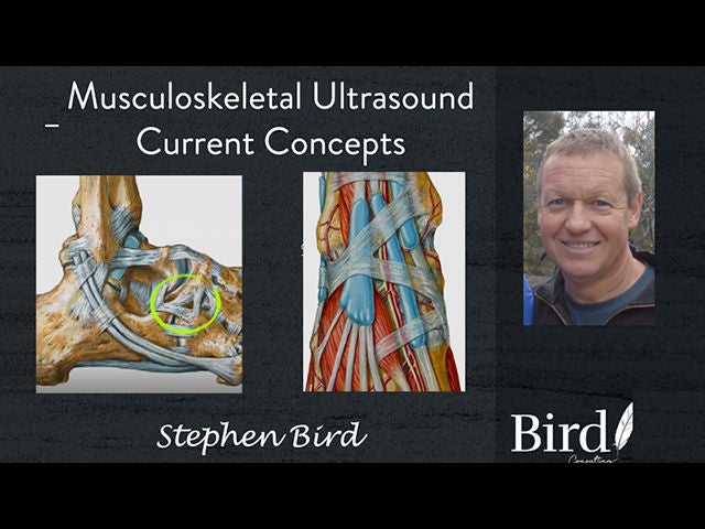
Ultrasound Clinical Case Study with Stephen Bird - Rheumatoid Elbow
Ultrasound Clinical Case Study with Stephen Bird - Elbow Radial Collateral Ligament
Ultrasound Clinical Case Study with Stephen Bird - Acute tendonosis - hydroxyapatite deposition
High Resolution Ultrasound in Genicular Nerve Radiofrequency Ablation
Schwannoma of the anterior cutaneous nerve
Clinical benefits and future prospects of ApliCam in the field of MSK
At the forefront of elite MSK ultrasound with the Aplio i800 / Prism Edition
The WOW-Effect in Sonography
High-resolution sonography redefines hand surgery diagnostics
Nerve Ultra sound Opens Up Completely New Perspectives
Guiding the Needle to the Deepest Layers of the Body
FHL effusion causing foot paraesthesia
Distal biceps tear
Radial collateral ligament tear
Lumbrical tear
Early adhesive capsulitis
Plantar plate tear
An indeterminate MRI for further assessment with Ultrasound
Schwannoma of the hand
Atheroma or Ganglion?
Seeking more information? We can help.

Social Media


