


See and deploy complex devices with confidence
New Alphenix Hi-Def Detector
Visualisation like never before with Canon Medical's first High-Definition (Hi-Def) mode*
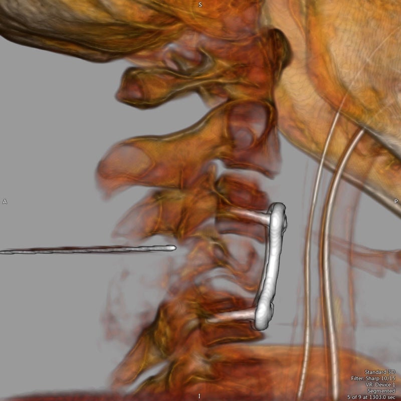
The Alphenix interventional systems feature the world’s first high definition (Hi-Def) detector with 76 micron pixel imaging mode. With more than twice the spatial resolution of conventional flat panel detectors (FPD), the Hi-Def detector helps clinicians visualise fine details and anatomical structures, and deploy devices with accuracy and confidence.
The unique Hi-Def imaging mode allows you to effortlessly zoom up to 4cm (1.5”) without losing image quality.
- Available as an option only
Hi-Def Clinical Gallery
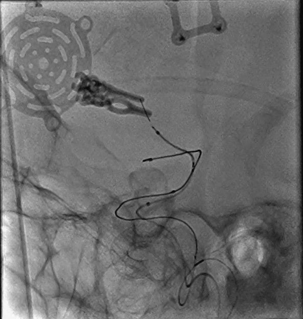
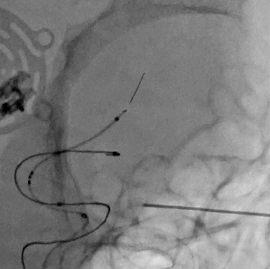
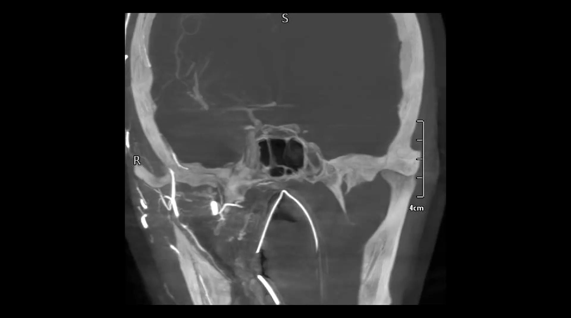
Stent-Assisted Coiling
Hi-Def imaging provides the highest possible resolution to accurately evaluate stent apposition. Incomplete stent apposition is a risk factor during stent-assisted coiling of wide-necked intracranial aneurysms and can result in a delayed ischemic event post treatment. Hi-Def imaging enhances clinical confidence and improves patient outcomes.



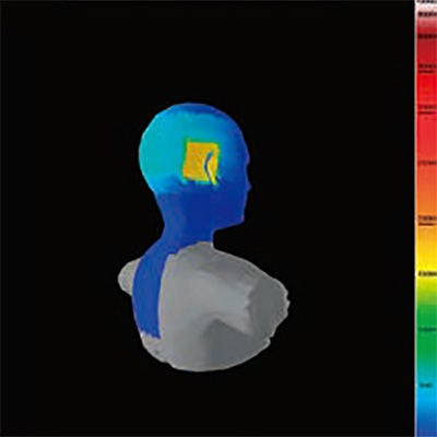
A study by The St George Hospital found that use of advanced Dose Reduction Technology (DTS) resulted in a 46% reduction in peak skin dose. Researchers confirmed that ongoing real-time dose feedback is a critical factor in the successful modification of operator behaviour to reduce radiation dose. *
- Wilson S, Prasan AM, Virdi A, et al. Real-time colour pictorial radiation monitoring during coronary angiography: effect on patient peak skin and total dose during coronary angiography. EuroIntervention 2016;12: 939–47.
DoseRite
Spot ROI – Up to 85% Reduction in dose
The unique Spot ROI collimator filter allows the operator to reduce dose to surrounding anatomy while simultaneously ensuring full image quality in the area that matters most.
Spot ROI:
- Can be positioned anywhere with the Field of View (FOV)
- Can be used to significantly reduce dose during fluoroscopic and DA runs
- Allows the operator to monitor the position of your guide wire and the surrounding anatomy, at significantly lower dose
DoseRite
Spot Fluoro - Reduce scatter radiation by up to 50% with advanced collimation
See a more complete picture with less dose to surrounding anatomy. Spot Fluoro enables the operator to collimate a rectangular or square region of interest (ROI) anywhere within the general field of view.
- Reduce cumulative dose area product by up to 50%**
- Reduce scatter radiation by more than 50%**, enabling safer procedures for patients and interventional staff
- Freely collimate anywhere on the image with asymmetric blade movements
- Last Image Hold (LIH) overlay with live fluoroscopy
- Next generation automatic brightness control with scatter correction, providing optimal image quality and clarity where you need it
- *Borota L, Jangland L, Aslund PE, et al. Spot fluoroscopy: a novel innovative approach to reduce radiation dose in neurointerventional procedures. Acta Radiol. 2017;58(5):600-608
Live Zoom
Image Magnification without Dose Penalty
- 50% dose reduction compared to geometric magnification
- Digitally magnify the image display without increasing detector input dose
- Increases image display size in real-time during both fluoroscopy and digital acquisition (DA,) offering dose savings compared to traditional FOV magnifications
13cm FOV - Fluoro
13cm FOV + Live Zoom 1.4 - Fluoro
Interventional Neuroradiology
Toolkit
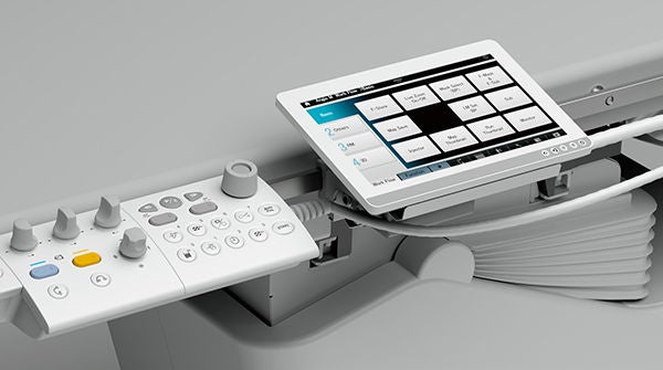
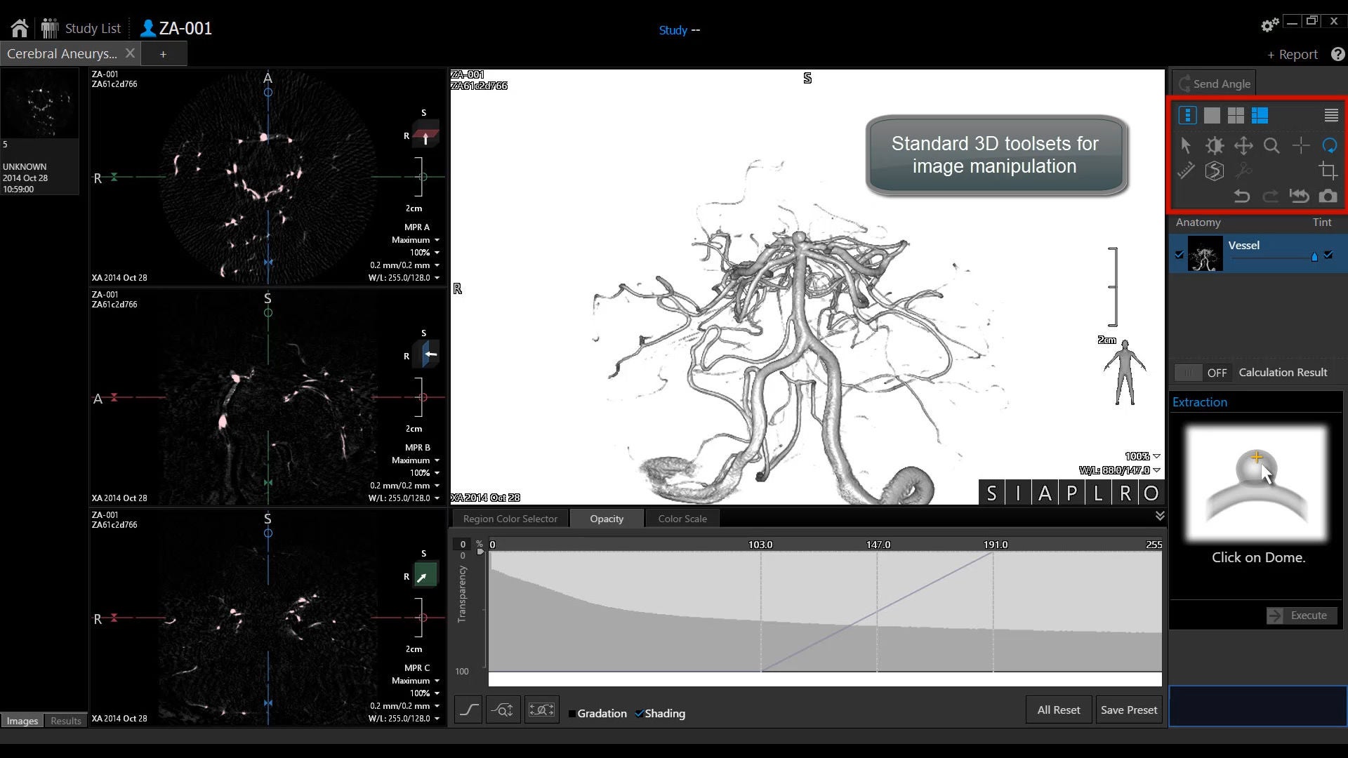
The Alphenix systems provide a comprehensive suite of advanced imaging tools to enhance visualisation. Applications are integrated into the new feature-rich workstation to improve workflow.
Advanced Interventional Software
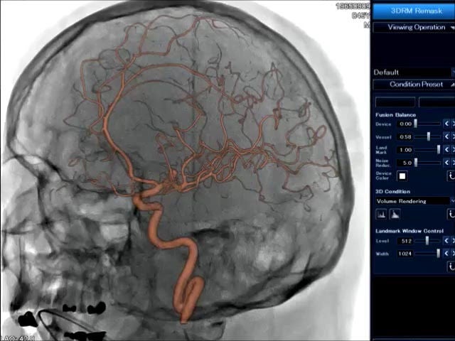
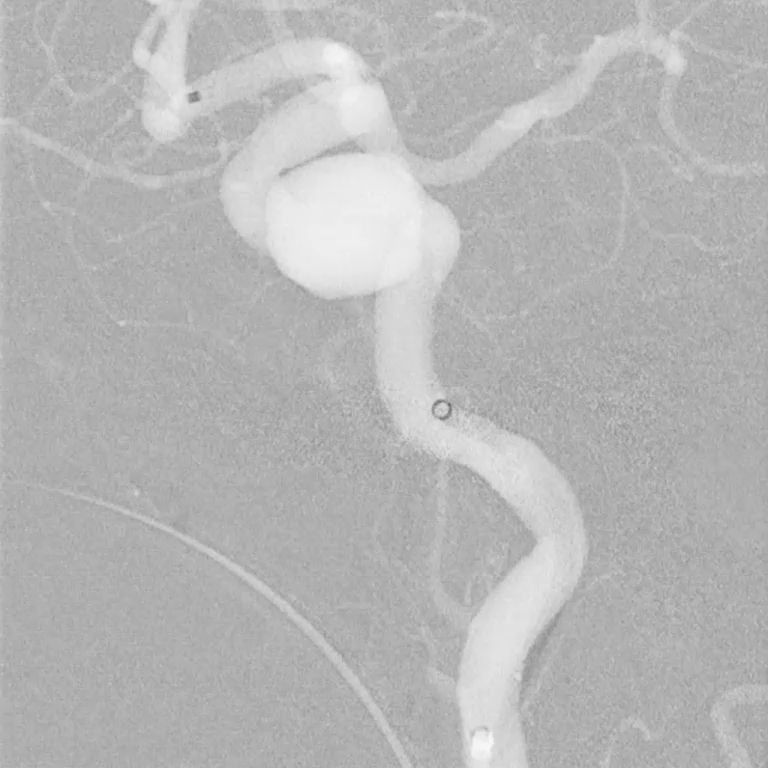
3D Angio
Fast and Intuitive Workflow
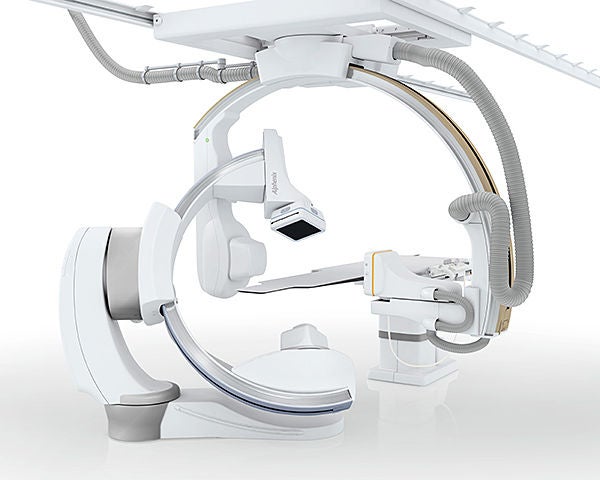
The fastest single plane, ceiling mounted C-arm available. The Alphenix allows rotations up to 80 deg/sec to provide faster 3D and cone beam CT times.
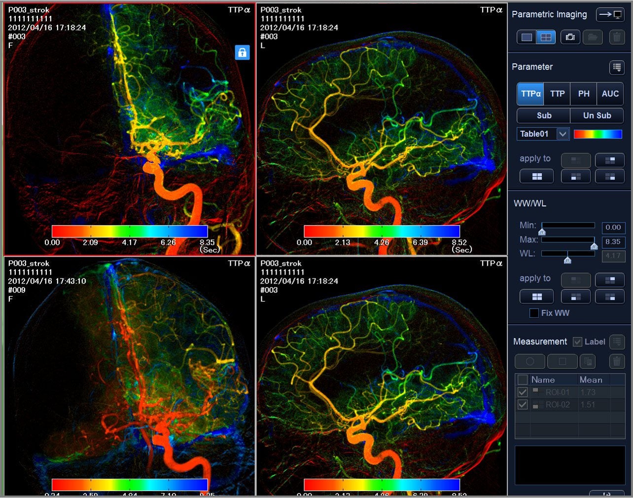
Parametric Imaging1
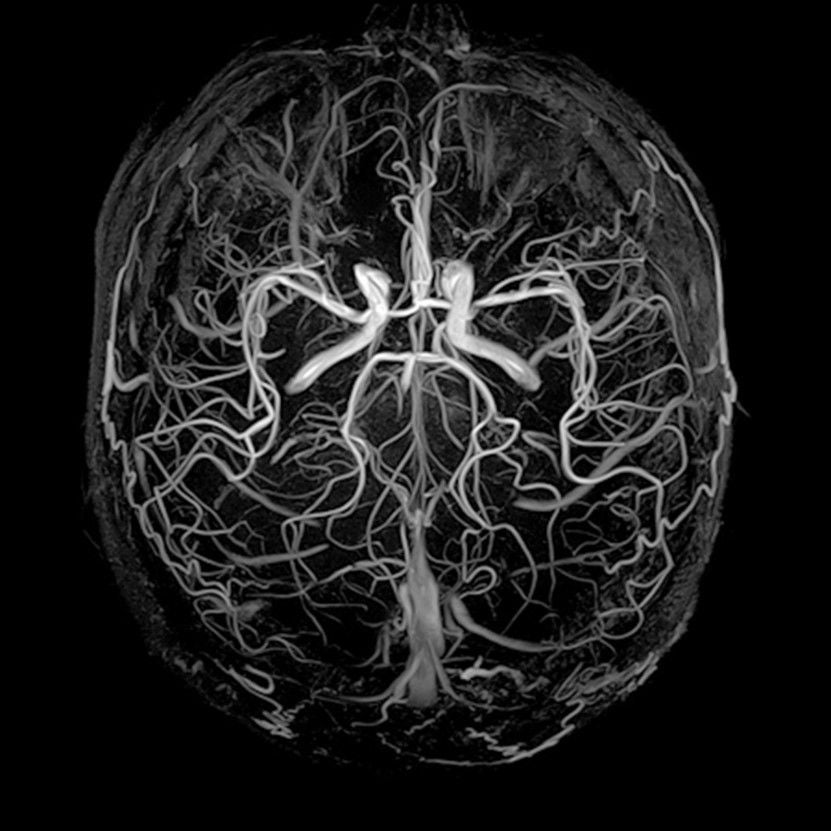
- Video playback with Colour Coded Circulation
- Displays an entire image sequence as a single composite image
- Colour coded in order to characterise contrast media dynamics and allow easier visual evaluation of patients
Parametric parameters include:
- Time To Peak (TTP)
- Time To Peak Alpha (TTPα)
- Peak Height (PH)
- Area Under Curve (AUC)
- Time To Arrival (TTA)
- Time To Arrival Alpha (TTAα)
- Mean Transit Time (MTT)
¹Parametric Imaging software is not intended for stand-alone use or diagnosis.
System Options
Flexible Configurations to Address Your Clinical Needs
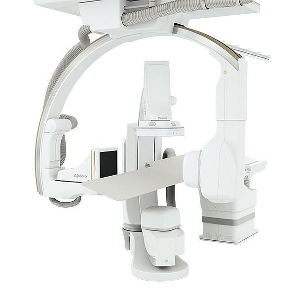
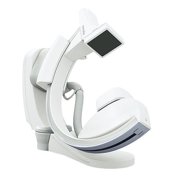
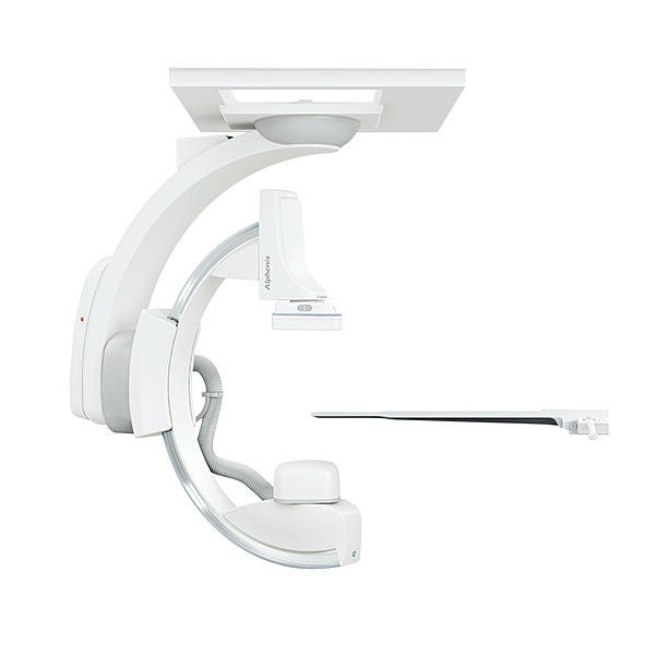
Developed in collaboration with leading clinicians, Alphenix offers a range of C-arm positioners designed to offer unprecedented access to your patient.

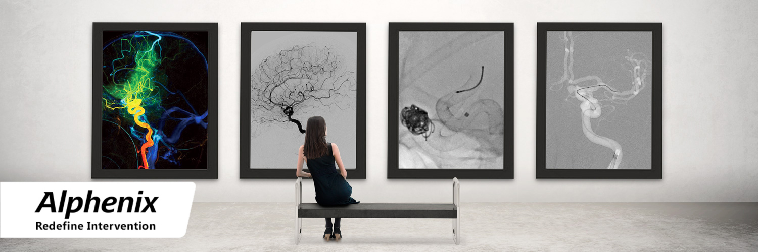
Alphenix Biplane Hi-Def: A Point Where Cutting-Edge Technologies and Fine Art Meet
The High Definition function offers not only excellent visualisation of the smallest anatomical structures and the smallest devices and their ultrastructure, but also offers the operator a unique visual, artistic experience.
- Dr. Ljubisa Borota, Uppsala University, Sweden
Neuroendovascular Treatment Using an Angiography System with the Hi-Def Detector
CAA includes a semiautomatic function that allows the user to perform setting and editing during neckline determination in cerebral aneurysm volume analysis. This is a very useful tool for formulating the optimal treatment strategy.
- Dr. Tetsu Satow, Chief Physician, Department of Neurosurgery
Benefits Using High-Definition (Hi-Def) Technology in Complex Neurovascular Procedures
The Hi-Def technology by Canon Medical, provides a new visualisation gateway to clinically expand the horizons of neuro intervention beyond the standard conventional imaging technology today.
- Adnan Siddiqui MD, PhD, FAHA
Experience using the New Alphenix Interventional System for Neuro-endovascular Therapy
During neurovascular therapy being able to see the intricate details in anatomy, and access devices gives us the confidence to quickly and better treat our patients with less chance of complications.
- Prof. Ichiro Nakahara, Department of Comprehensive Strokology, Fujita Health University Hospital
Initial investigations of a special high-definition (Hi-Def) zoom capability in a new detector system for neuro-interventional procedures
The Hi-Def zoomed images were much preferred over the FPD, indicating the potential to improve endovascular procedures and hence outcomes using such a Hi-Def feature.
S.V. Setlur Nagesh et al, NCBI
Quantitative comparison of Hi-Def and FPD zoom modes in an innovative detector using Relative Object Detectability (ROD) metrics
Due to their higher resolution, high-definition-mode images are sharper and visually preferred compared with the flat panel detector images. The improved imaging provided by the high-definition mode can potentially provide an advantage during neurointerventional procedures.
J. Krebs et al, SPIE Digital Library
High-Definition Zoom Mode, a High-Resolution X-Ray Microscope for Neurointerventional Treatment Procedures: A Blinded-Rater Clinical-Utility Study
Each of the metrics demonstrated improved performance of the Hi-Def zoom mode over the standard FPD mode in imaging a wide array of objects such as stents, wires, and spheres. Of particular note is the greater performance of the Hi-Def when considering the high spatial frequencies necessary for visualising fine image details of a pipeline stent.
S.V. Setlur Nagesh et al, American Journal of Neuroradiology
Refine and Redefine Intervention Using High-Definition (Hi-Def) Technology
Andrew Kuhls-Gilcrist, PhD, DABR, Mark Hohn, Dale Marek, RT(R), Yiemeng Hoi, PhD Medical Affairs, Interventional X-ray, Canon Medical Systems USA, Inc.
Swetadri Vasan Setlur Nagesh, PhD, Stephen Rudin, PhD, FAAPM, Adnan Siddiqui, MD, PhD, FACS, FAHA, FAANS Canon Stroke and Vascular Research Centre University at Buffalo, Buffalo, NY
High Definition Imaging Reduces Procedure Time Without Impacting Patient Dose in Image-Guided Neuro Interventional Procedures.
Swetadri Vasan Setlur Nagesh, MS,PhD, Buffalo, NY (Presenter) Nothing to Disclose
Single-centre experience of using high definition (Hi-Def) imaging during neurointervention treatment of intracranial aneurysms using flow diverters.
PSwetadri Vasan Setlur Nagesh1,2, Kunal Vakharia2,3, Muhammad Waqas2,3, Stephan A Munich2,3, Daniel R Bednarek1,2,4, Jason M Davies2,3,5, Kenneth V Snyder2,3, Maxim Mokin6, Stephen Rudin1,2,4,7,8,9, Elad I Levy1,2,3,4, Adnan H Siddiqui 1,2,3,4
Social Media


