

Ultrasound Clinical Case Study with Stephen Bird
Elbow Radial Collateral Ligament

Stephen Bird
Sonographer – Benson Radiology
AMS, AMS (Vascular), DMU, DMU (Vascular), MMedSon
A general sonographer with 25 years clinical experience, Stephen has a strong commitment to advancing ultrasound education. In 2002 Stephen received the ASA Pru Pratten Memorial Sonographer Achievement Award and in 2008 he was made an Honorary Fellow of ASUM.
Learn more about Stephen Bird's online courses and webinars.
AMS, AMS (Vascular), DMU, DMU (Vascular), MMedSon
A general sonographer with 25 years clinical experience, Stephen has a strong commitment to advancing ultrasound education. In 2002 Stephen received the ASA Pru Pratten Memorial Sonographer Achievement Award and in 2008 he was made an Honorary Fellow of ASUM.
Learn more about Stephen Bird's online courses and webinars.
Introduction
The radial collateral ligament (RCL) is a strong, short, thin band of tissue that fans out from the anterolateral aspect of the radiocapitellar joint. It runs longitudinally underneath the common extensor tendon, blending with the anterior annular ligament, and its posterior fibres merge with the lateral ulnar collateral ligament.
The primary role of the RCL is to act as a static stabiliser, providing stability against varus force to the joint.
Ultrasound is a valuable tool in the assessment of patients with suspected tendinosis of the common extensor origin (CEO), otherwise referred to as a tennis elbow. It is common to observe a classic pattern of disrepair phase tendinosis affecting primarily the extensor carpi radialis brevis (ECRB) tendon origin. The tendon appears hypoechoic, and high-sensitivity Doppler algorithms such as Canon’s Superb Microvascular Imaging (SMI) or Advanced Dynamic Flow (ADF) are effective at demonstrating associated hypervascularity.
Like all large synovial joints, the elbow has a capsule constructed of collagen, and the various load bearing components of the joint capsule are strategically thickened becoming capsular ligaments. Deep to the CEO lies the RCL and annular ligament components of the joint capsule. Synovial fluid should be contained within the joint by the capsular ligaments ensuring no fluid is present directly beneath the common extensor origin.
Case study
This case features a retired 75-year-old female with a history of physically demanding employment. The patient presented with chronic lateral elbow pain that was unresponsive to oral anti-inflammatory treatment and an unguided steroid injection.
An ultrasound examination was performed on the Canon Medial Aplio a550 using a high frequency linear transducer (18L7).
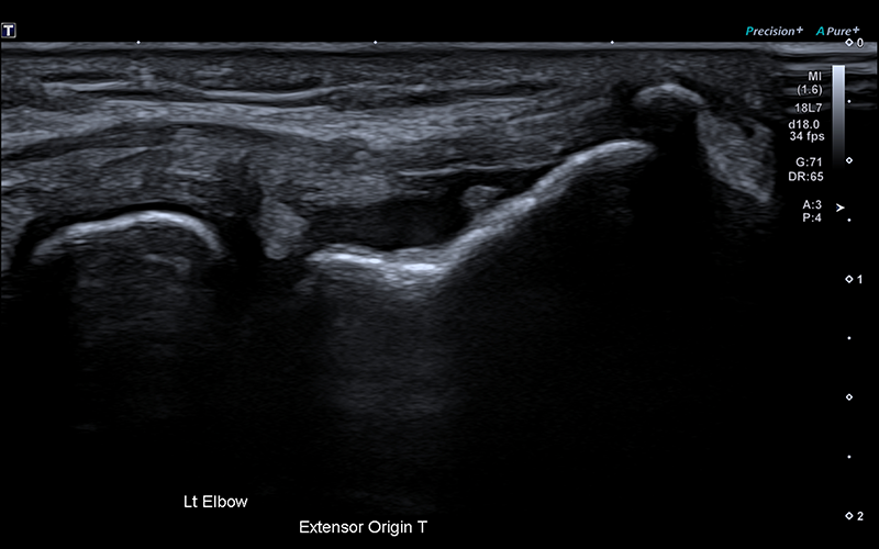
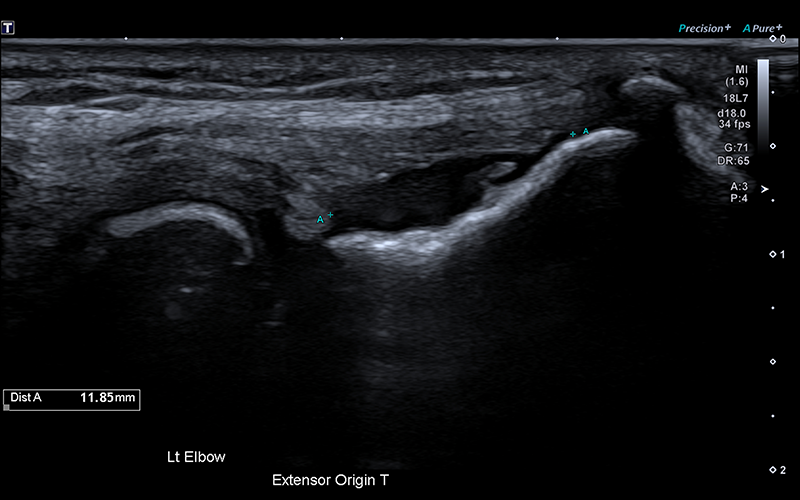
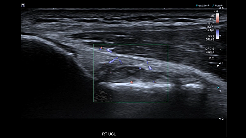
Long axis imaging of the patients CEO clearly demonstrates joint fluid flowing freely between the radial head and humeral capitellum (Figure 1). The retracted free end of the RCL is visualised hanging like a tear drop of echogenic collagen into the synovial fluid. The length of the joint capsule defect can be estimated by measuring the distance between the retracted ‘tear drop’ end of the RCL at its normal original location on the lateral epicondyle (Figure 2).
Discussion
Short axis imaging can be extremely valuable in assessing the CEO. In the short axis it is very easy to localise the ECRB component of the CEO from the muscle belly and this allows the sonographer to follow the tendon proximally as they slide the transducer over the radial head and onto the lateral epicondyle. In this plane, it is essential to observe a continuous line of joint capsule collagen deep to the CEO, with the synovial fluid beneath it.
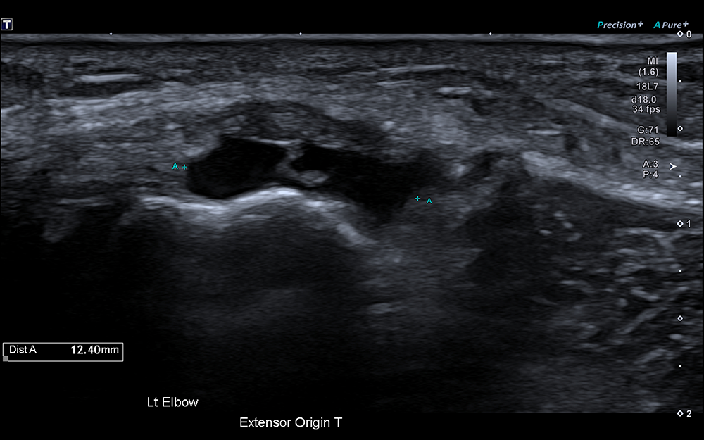
In this clinical case high-resolution, short axis imaging provided clear visualisation of the defect in the joint capsule at the level of the lateral epicondyle and allowed the width of the joint capsule tear to be measured (Figure 3).
High-resolution imaging with fewer artifacts is essential for complex MSK examinations. The Aplio a550 integrates industry leading imaging technologies to deliver exceptional detail to diagnose quickly with ease and confidence. The system’s unique aBeam architecture ensure all the unique imaging technologies work seamlessly together to provide greater uniformity, enhanced penetration and improved visualisation. The system’s lightweight transducers feature outstanding clinical versatility, ergonomic construction and thin, light-weight cables, designed to increase operator efficiency
Other Aplio a550 Case Studies by Stephen Bird
Social Media


