

Ultrasound Clinical Case Study with Stephen Bird
Rheumatoid Elbow

Sonographer – Benson Radiology
AMS, AMS (Vascular), DMU, DMU (Vascular), MMedSon
A general sonographer with 25 years clinical experience, Stephen has a strong commitment to advancing ultrasound education. In 2002 Stephen received the ASA Pru Pratten Memorial Sonographer Achievement Award and in 2008 he was made an Honorary Fellow of ASUM.
Learn more about Stephen Bird's online courses and webinars.
AMS, AMS (Vascular), DMU, DMU (Vascular), MMedSon
A general sonographer with 25 years clinical experience, Stephen has a strong commitment to advancing ultrasound education. In 2002 Stephen received the ASA Pru Pratten Memorial Sonographer Achievement Award and in 2008 he was made an Honorary Fellow of ASUM.
Learn more about Stephen Bird's online courses and webinars.
Introduction
Clinical diagnosis of synovial proliferative diseases is challenging because symptoms are often non-specific (1). Advanced imaging such as high-resolution ultrasound can aid in revealing characteristic findings of synovial proliferative diseases, which is key to an accurate diagnosis and for early treatment (2).
Synovial diseases can be inflammatory, infectious, neoplastic, degenerative, traumatic or haemorrhagic in nature (2). They can be a part of a systemic disease, such as (inflammatory) arthritis, or a primary process in a single joint. Bone erosions typically occur as part of a pathologic process involving the synovium and can result in irreversible joint destruction.
Articular bone erosions are now recognised as a central finding in rheumatoid arthritis and can be associated with poor functional outcomes (3). When synovial inflammation develops from autoimmunity, additional triggers augment the process of bone erosion.
Synovial diseases can be inflammatory, infectious, neoplastic, degenerative, traumatic or haemorrhagic in nature (2). They can be a part of a systemic disease, such as (inflammatory) arthritis, or a primary process in a single joint. Bone erosions typically occur as part of a pathologic process involving the synovium and can result in irreversible joint destruction.
Articular bone erosions are now recognised as a central finding in rheumatoid arthritis and can be associated with poor functional outcomes (3). When synovial inflammation develops from autoimmunity, additional triggers augment the process of bone erosion.
Case study
This patient presented with diffuse elbow pain, which had increased in intensity over several months with no history of trauma. The pain was poorly localised, and the patient indicated that symptoms were affecting all aspects of the joint. This suggested that that the pathology was most likely joint related, rather than related to a tendon or neurological in nature.
An ultrasound examination was performed on the Canon Medial Aplio a550 using the 14L5 and high frequency 18L7 linear transducers.
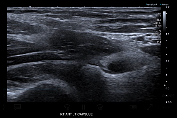
On review of the coronoid and annular recesses of the anterior elbow joint, a significant joint effusion with displacement of the anterior fat pad and convexity of the annular ligament is evident (video 1). Closer analysis also revealed considerable synovial proliferation of the elbow joint capsule.
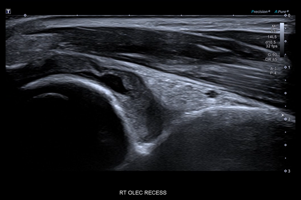
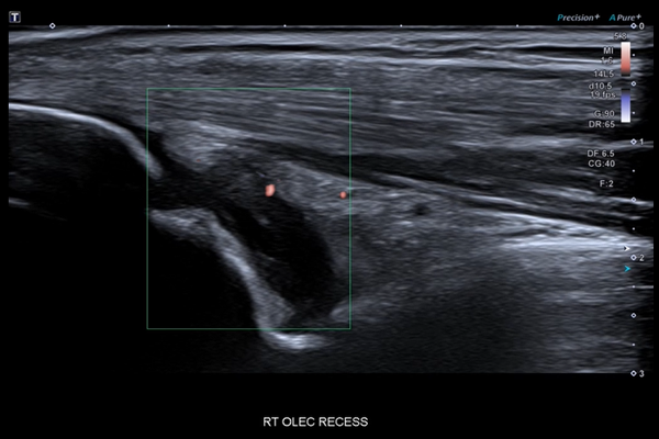
Video 2 shows the olecranon recess of the elbow joint deep to the triceps apparatus. A joint effusion is clearly evident, accompanied by significant synovial hypertrophy. With Canon’s Advanced Dynamic Flow (ADF) the synovial component of the joint capsule came to life with vascularity (video 3).
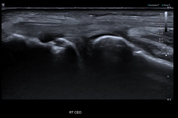
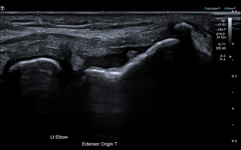
Minor structural changes in the common extensor origin were observed, suggesting tendinosis (video 4). However, minimal flow was demonstrated with ADF, indicating that these changes were unlikely contributing to the patient’s pain. Deep to the common extensor origin, the radial collateral ligament and annular ligament were hypoechoic, thick and ADF assessment elicited significant hypervascularity (video 5). Bone erosion at the lateral epicondyle of the humerus is clearly visible, providing another clinically significant indication.
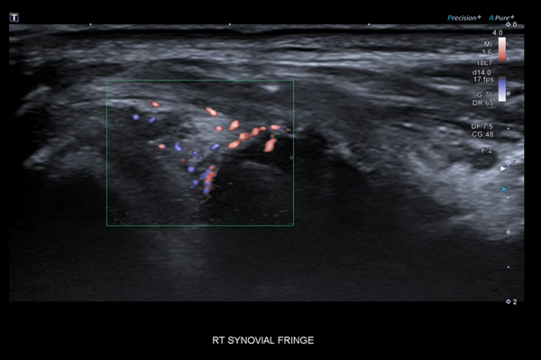
Video 6 shows the common flexor origin and the anterior band of the ulnar collateral ligament. Immediately deep to the ulnar collateral ligament is the intra-articular space of the elbow joint, which normally contains a small amount of fat and synovial fluid. ADF demonstrated hypervascularity indicating synovial hypertrophy (video 7).
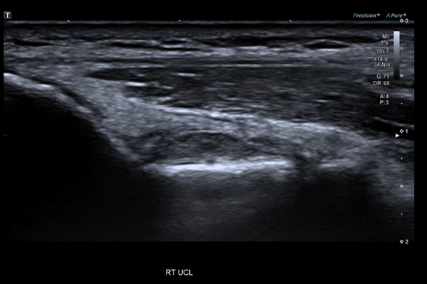
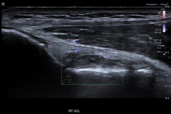
The constellation of findings indicates synovial proliferative disease with associated bone erosions. This likely signifies rheumatoid disease and a rheumatological work up was recommended.
Discussion
Advanced Dynamic Flow played a key role in many of the main findings of this case. Available on all Canon Medical Aplio ultrasound systems, ADF offers high-resolution colour flow that enables physicians to quickly identify small complex vascularity.
The Aplio a550 integrates industry-leading imaging technologies to deliver depth and detail to diagnose quickly with ease and confidence.
References
1. Synovial Tumors and Proliferative Diseases [Read here]
2. Basic radiological assessment of synovial diseases: a pictorial essay.
Aynur Turan, Pınar Çeltikçi, Abdurrahman Tufan, and Mehmet Akif Öztürk [Read here]
3. Bone erosion in rheumatoid arthritis: mechanisms, diagnosis and treatment
Georg Schett and Ellen Gravallese [Read here]
Social Media


