Search

Image

Alphenix solutions for interventional radiology and oncology

As interventional procedures and oncology treatments increase in complexity, Alphenix flexibility supports you in delivering the best possible outcome to your patients.

image

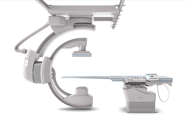
Move the system, not the patient

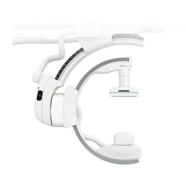
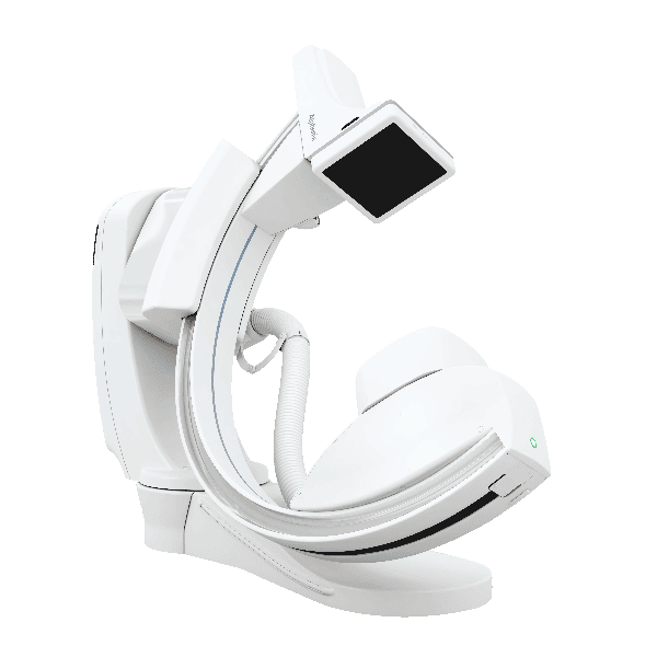
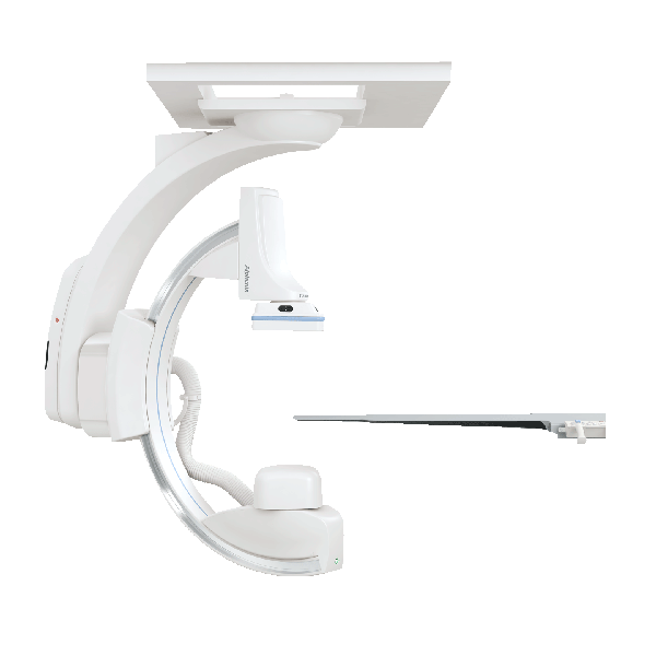
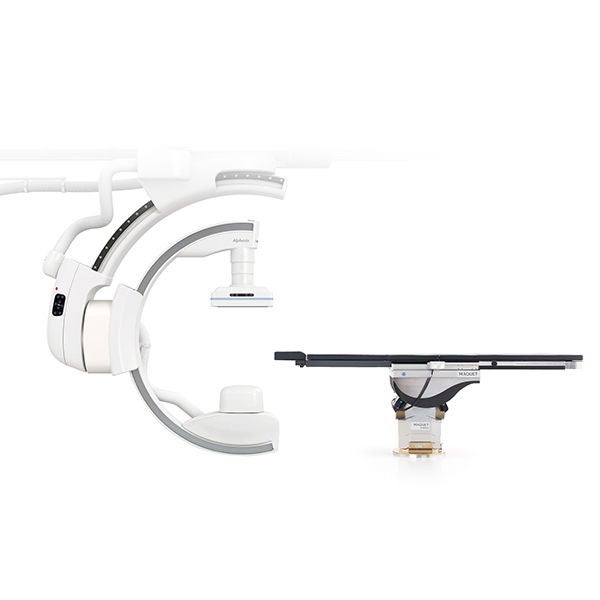
The Alphenix’s ceiling-mounted C-arm offers an unprecedented range of movement, allowing you to position the C-arm to suit your interventional workflow.

  • Full body coverage as standard 
  • Head end access - providing improved workflow and patient access 
  • Tableside 3D capability 
  • Fastest single plane ceiling-mounted C-arm available allowing rotations at up to 80 degrees per second, providing faster 3D and cone beam CT times.

WorkRite

Enhanced workflow to give you greater control and flexibility

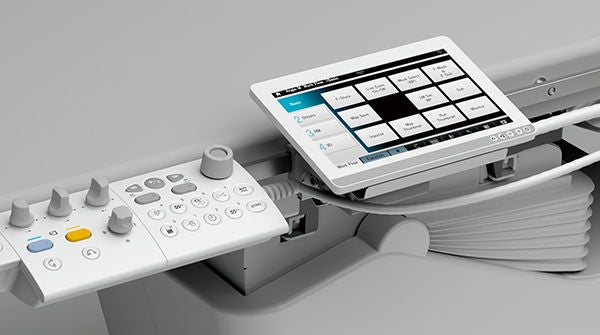
The new tableside Alphenix Tablet gives you control right at your fingertips to deliver a faster and more seamless workflow.

image

WorkRite

Smoother procedures start with better planning

The new feature-rich Alphenix Workstation better integrates applications to help plan, analyse and perform interventional procedures.

Interventional radiology features include embolisation planning, multi-modality fusion and coronary artery stent analysis.

image
image

Refine and redefine intervention using the world’s first High-Definition technology

The Alphenix interventional systems feature the world’s first high definition (Hi-Def)* detector with 76 micron pixel imaging mode. With more than twice the spatial resolution of conventional flat panel detectors (FPD), the Hi-Def detector helps clinicians visualise fine details and anatomical structures and deploy devices with accuracy and confidence.

*Available as an option only

Initial experience using a novel high definition (Hi-Def) imaging system in peripheral arterial interventions

“Our initial experience using a novel Hi-Def imaging system shows that it can effectively be used in real-time to improve visualisation of lower extremity vasculature and interventional devices during complex peripheral arterial interventions and may be used to improve technical outcomes.”

-Salman A. Arain, Steven Napierkowski, Michael Schechter, Wahaj Aman, Richard W. Smalling
 The University of Texas Health Science Center at Houston, Houston, TX

Alphenix Hi-Def Detector

Peripheral Intervention


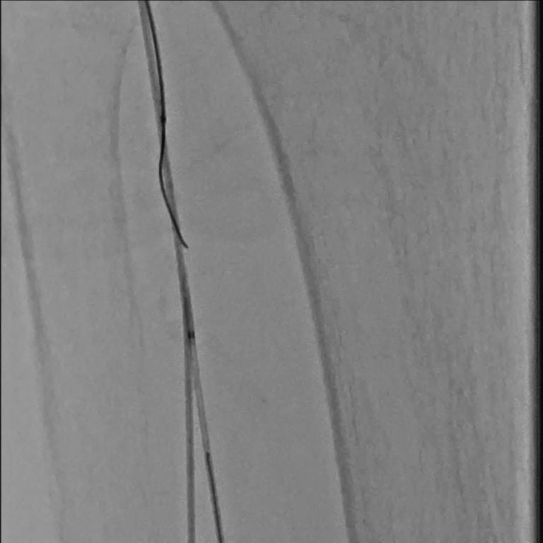
Hi-Def imaging is a valuable tool in challenging peripheral interventional cases.

With twice the spatial resolution of conventional FPD technology, Hi-Def mode can provide clinicians with the improved visualisation needed to pass guide-wires and engage catheters during challenging retrograde and antegrade revascularisation.
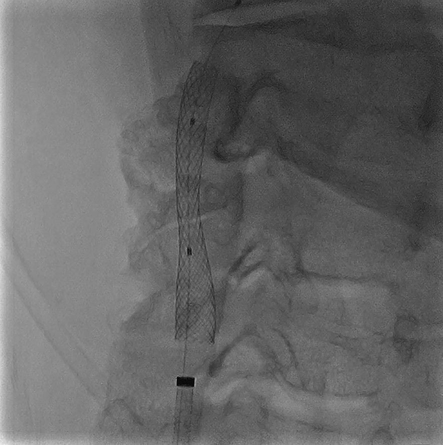
Carotid Stenting


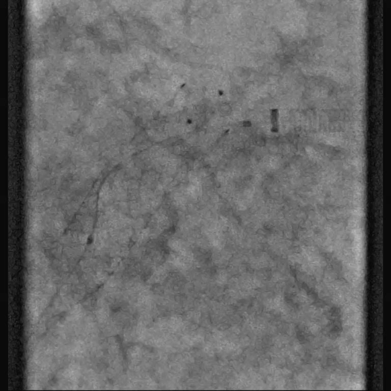
Hi-Def mode providing enhanced visualisation during the treatment of a symptomatic right ICA stenosis.

8.0 x 21mm WALLSTENT, 7.2mm Emboshield NAV6 embolic protection device and AVIATOR Plus 4.0 x 20mm balloon.
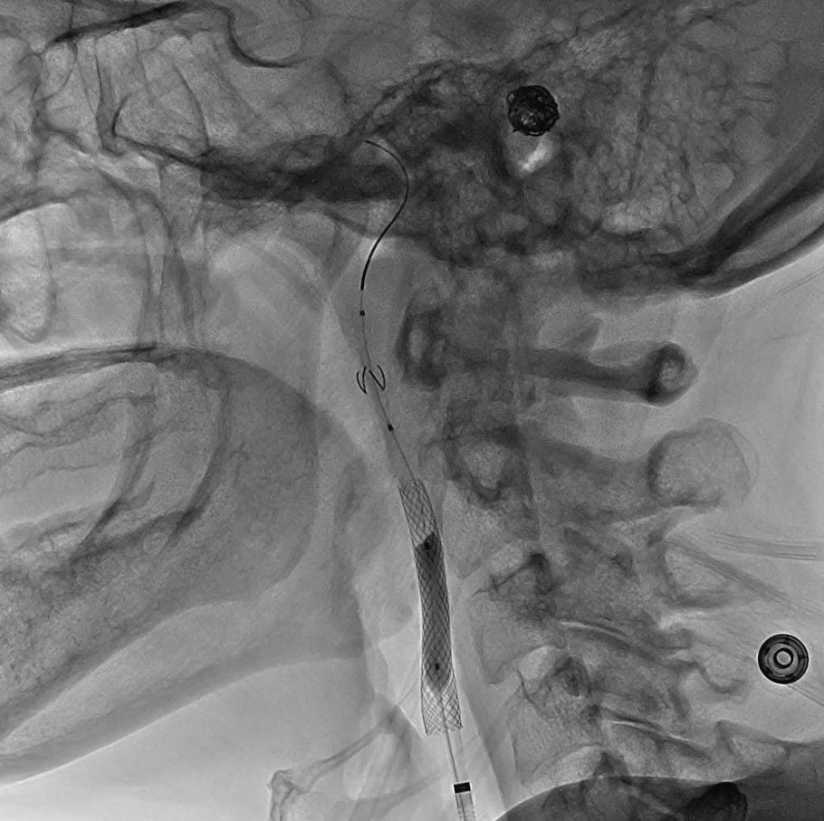
Lateral (8" Standard FOV)

image

Frontal (3" Hi-Def Zoom)

image
image

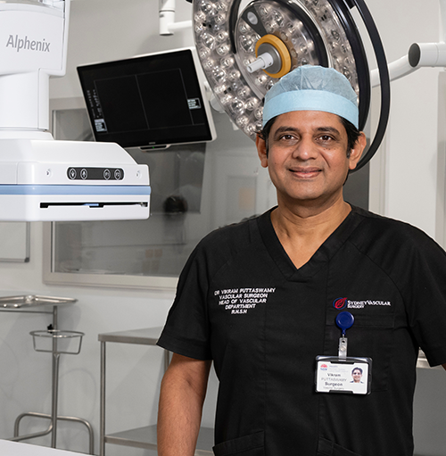
“Alphenix gives us exceptional spatial resolution and clarity. As the imaging gets clearer, our ability to do procedures with even more confidence increases.”

Dr. Vikram Puttaswamy
Vascular and renal transplant Surgeon
Royal North Shore Hospital
ImagingRite

Navigate with confidence and accuracy

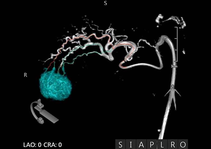
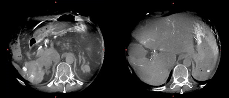
Comprehensive planning tools allow for quick and intuitive analysis prior to embolisation procedures. Using 3D volume data to select the tumor and define the corresponding feeding vessels, this information as an overlay can be viewed during live fluoro of the procedure. In multi-phase Alpha CT, visualisation of contrast media corresponds to arterial, venal and capillary blood flow providing valuable insight in identification of hepatic tumor feeding vessels.

ImagingRite

Enhanced visualisation during interventional procedures

An improved imaging chain with new hardware and software technologies provide you with fast and assured visualisation. Superimpose 3D volume data on the live fluoro display to provide additional views of the vascular anatomy to aid clinicians during interventional procedures.

image
image

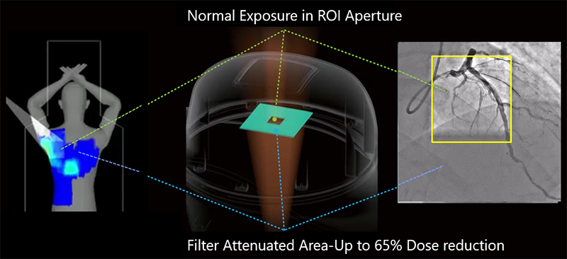
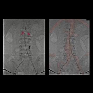
SpotROI - See more of interest, for less dose

The unique Spot ROI collimator filter allows the operator to reduce dose to surrounding anatomy while simultaneously ensuring full image quality in the area that matters most. This allows operators to always position the ROI over the vascular structure of interest, independently of its location with the FOV.

Spot ROI:

  • Can be positioned anywhere with the Field of View (FOV) 
  • Can be used to significantly reduce dose during fluoroscopic and DA runs 
  • Allows the operator to monitor the position of your guidewire and the surrounding anatomy, at significantly lower dose

DoseRite

Spot Fluoro - Reduce scatter radiation by up to 50% with advanced collimation

See a more complete picture with less dose to surrounding anatomy. Spot Fluoro enables the operator to collimate a rectangular or square region of interest (ROI) anywhere within the general field of view. Reduce cumulative dose area product by up to 50%**

  • Reduce scatter radiation by more than 50%**, enabling safer procedures for patients and interventional staff   
  • Freely collimate anywhere on the image with asymmetric blade movements    
  • Last Image Hold (LIH) overlay with live fluoroscopy    
  • Next generation automatic brightness control with scatter correction, providing optimal image quality and clarity where you need it

**Borota L, Jangland L, Aslund PE, et al. Spot fluoroscopy: a novel innovative approach to reduce radiation dose in neurointerventional procedures. Acta Radiol. 2017;58(5):600-608

DoseRite

Dose Tracking System - Visualise skin dose in real-time

DTS is a patient-centric model that records the patient’s skin dose in real-time according to imaging parameters and geometry to provide an accurate demonstration of the dose received during and examination.

Real-time dose monitoring and visualisation allows the operator to modify their approach during procedures to avoid regions where dose thresholds could be exceeded.


A study by The St George Hospital found that use of advanced Dose Reduction Technology (DTS) resulted in a 46% reduction in peak skin dose during coronary stent insertion. Researchers confirmed that ongoing real-time dose feedback is a critical factor in the successful modification of operator behaviour to reduce radiation dose. *

*Wilson S, Prasan AM, Virdi A, et al. Real-time colour pictorial radiation monitoring during coronary angiography: effect on patient peak skin and total dose during coronary angiography. EuroIntervention 2016;12: 939–47.

Interventional Radiology/Oncology

Toolkit

The new feature-rich Alphenix Workstation better integrates applications to help you plan, analyse and perform procedures.

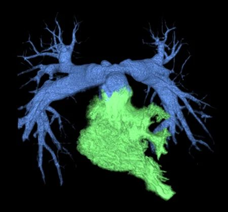
⁠3D Angio

Fast and Intuitive Workflow

3D rotational speed up to 50 deg/sec
image

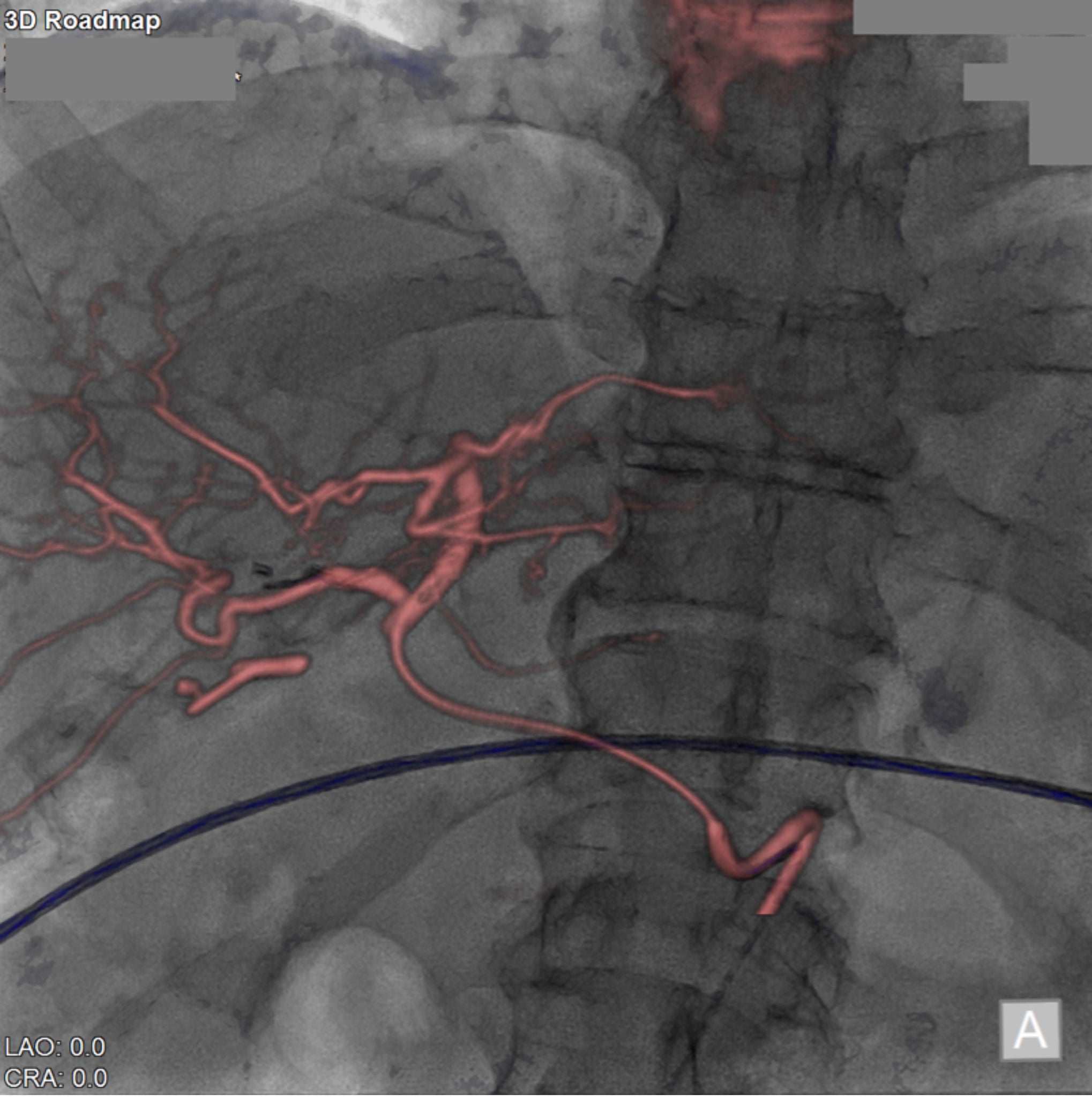
3D Roadmap

Turn 3D Acquisitions into Live Fluoro Roadmaps

3D image display changes according to the system conditions such as the C-arm angle, SID, field size, tabletop height, and tabletop panning.

Image

Alpha CT

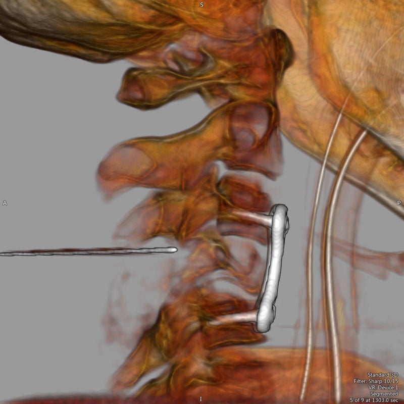
To supplement 3D imaging, CT-like Imaging is available to support visualisation of anatomy or pathology during interventional procedures.

Up to 3 CBCT continuous acquisitions phases are available to support the planning for TACE (Transcatheter Arterial Chemoembolisation)


• Head         • Neck
• Body         • Device
• Vessel       • MAR
image

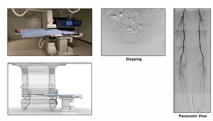
⁠DSA Stepping

  • Used in lower extremity 
  • Takes a mask of abdomen to feet 
  • Second run allows for the table to be moved (stepped) 
  • The clinician follows the injection of contrast from the aorta to the arteries of the feet

Dynamic Trace

Can be used in combination with DSA or to supplement DSA or for stand-alone imaging Dynamic Trace reduces the effects of the bones and enhances background compression to emphasize the blood vessels.

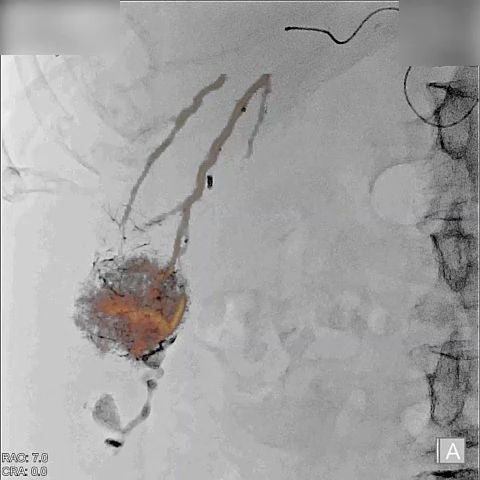
 Embolisation Plan

Rapid tumour and feeder vessel extraction

  • Identify and extract up to ten tumours and associated feeder vessels using the Alphenix Workstation 
  • Utilise CTA or Alpha CT datasets 
  • Overlay with Fluoro utilising 3D Roadmap (Alpha CT) or Multi-Modality Fusion (CTA)
Multi-Modality Fusion

Enhanced visualisation during interventional procedures

Canon Medical's roadmap function enables 3D CT or MR Volume data to be superimposed on the live fluoro display. This feature provides additional views of the vascular anatomy to aid clinicians during interventional

image

Needle Guidance

CT or XA Datasets

Needle guidance function enables positioning of the C-arm to the optimum viewing position and display of a puncture guide line for assistance.

image

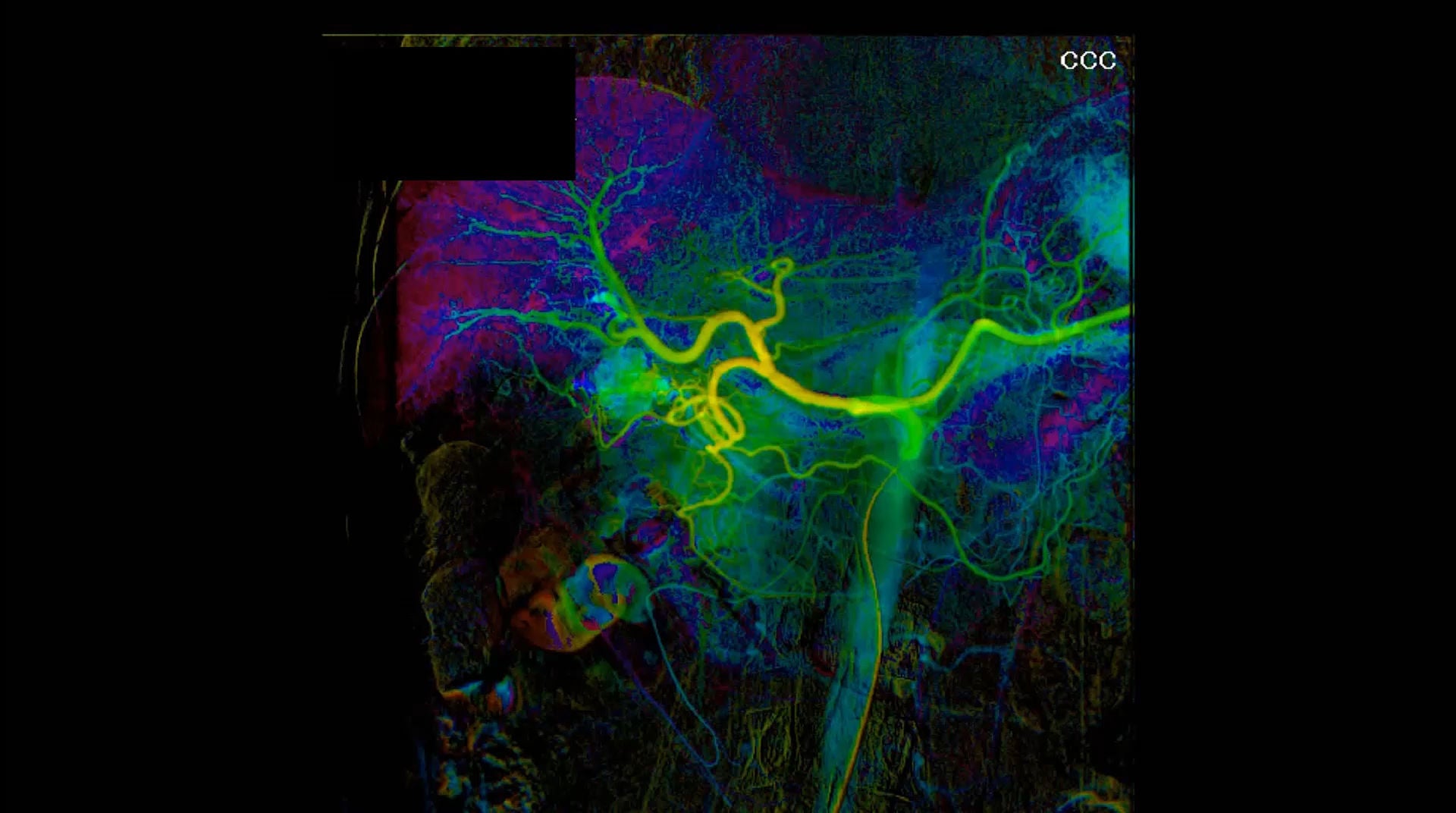
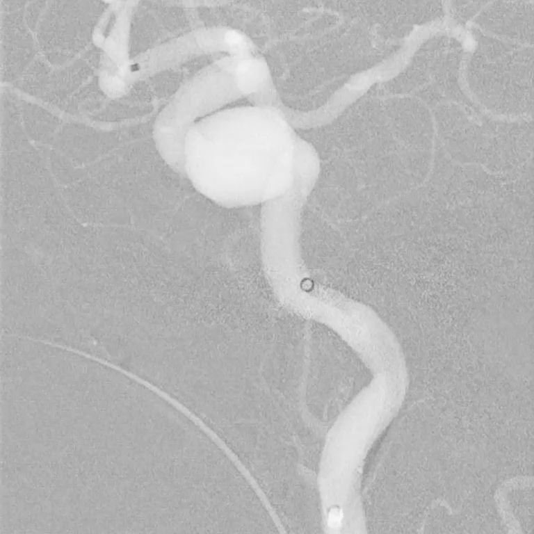
Parametric Imaging¹

  • Video playback with Color Coded Circulation 
  • Displays an entire image sequence as a single composite image 
  • Color coded in order to characterise contrast media dynamics and allow easier visual evaluation of patients

¹Parametric Imaging software is not intended for stand-alone use or diagnosis.


Parametric parameters include:


•Time To Peak (TTP)                  • Time To Peak Alpha (TTPα)
• Peak Height (PH)                    • Area Under Curve (AUC)
• Time To Arrival (TTA)              • Time To Arrival Alpha (TTAα)
• Mean Transit Time (MTT)
⁠Instant Roadmap

Auto Peak Contrast Detection

Automatic image adjustment during fluoro.

Image

System Options

Flexible Configurations to Address Your Clinical Needs
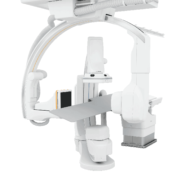
Alphenix Biplane

(Multi-access biplane system)

Combines the exception flexibility of a floor-mounted and ceiling-mounted C-arm combination, the biplane system is the ideal choice in complex interventional procedures.

Image

Alphenix Sky +

(Advanced ceiling-mounted system)

Advanced ceiling-mounted C-arm offers unprecedented flexibility and full body 3D imaging capability with faster acquisition, fewer artifacts and less contrast agent.

Image

Alphenix Core +

(Floor-mounted multi-access single-plane system)

Provides unmatched flexible patient access, the five-axis floor-mounted C-arm is ideally suited for a wide range of interventional applications.

Image

Alphenix Sky

(Ceiling-mounted)

Features a unique ceiling-mounted C-arm to provide longitudinal and lateral coverage to support upper extremity examinations.

Image

Alphenix Hybrid +

(Advanced ceiling-mounted system integrated with hybrid OR)

Dedicated for use with an operating table, advanced ceiling-mounted C-arm offers unprecedented flexibility and full body 3D imaging capability with faster acquisition, fewer artifacts and less contrast agent.

Image

System Options

⁠Configure your Alphenix Hybrid Angio-CT to meet your needs.


Alphenix Sky +
Advanced ceiling-mounted C-arm offers unprecedented flexibility and full body 3D imaging capability with faster acquisition, fewer artifacts and less contrast agent.

Aquillion ONE / INSIGHT Edition
(80 cm bore, 640 slices*, 16cm/rotation volume scan coverage)
For further information, please click here.


Alphenix Sky +
Advanced ceiling-mounted C-arm offers unprecedented flexibility and full body 3D imaging capability with faster acquisition, fewer artifacts and less contrast agent.

Aquillion Exceed LB
(90 cm bore, 160 slices*)
For further information, please click here.


Alphenix Sky +
Advanced ceiling-mounted C-arm offers unprecedented flexibility and full body 3D imaging capability with faster acquisition, fewer artifacts and less contrast agent.

Aquilion ONE / GENESIS Edition
(78 cm bore, 640 slices*, 16cm/rotation volume scan coverage)
For further information, please click here.


Alphenix Sky
Features a unique ceiling-mounted C-arm to provide longitudinal and lateral coverage to support upper extremity examinations.

Aquilion Prime SP
(78 cm bore, 160 slices*)
For further information, please click here.

Refine and redefine intervention using high-definition (Hi-Def) technology

Average procedure time, average fluoro time, the total number of X-ray pulses and cumulative air kerma were all significantly reduced …

Andrew Kuhls-Gilcrist et al


Expanding Interventional Radiology with the Highest Image Quality

“The system is really addictive. I cannot think how to treat my patients without the Alphenix 4D CT.”
Prof. Franco Orsi, Head of the Interventional Radiology department, IEO, Milan, Italy.

Sky’s the Limit for Royal Bournemouth with Alphenix Sky Ceiling-Mounted System

“The system has been really intuitive to use and we have received really good apps support from the Canon Medical team.”

Anne-Marie Jenkins, X-ray and Ultrasound Service Manager at Royal Bournemouth Hospital

Alphenix 4D CT – Cutting-Edge Innovation in one Single Room

“With good results and low morbidity the techniques possible with the Alphenix 4D CT system provide high-quality patient care at low medical cost.”
Prof. Alban Denys, Head of Interventional Radiology at CHUV.

The Alphenix 4D CT – More Treatments in a More Accurate Manner

“With Canon’s new Angio CT system, we have become more efficient and have been able to shorten the waiting times significantly for several interventions.”
Dr. Olle Haller, Head of Interventional Radiology, Gävle Central Hospital, Sweden.

Maximum Efficiency, Prioritized Patient Safety and Increased Revenue with Canon Medical’s 4D CT Technology

Osman Ahmed, MD Section of Vascular and Interventional Radiology Department of Radiology University of Chicago Medical Center

Yiemeng Hoi, PhD, Dale Marek, RT(R), Mark Hohn, Andrew Kuhls-Gilcrist, PhD, DABR Medical Affairs, Interventional X-ray Canon Medical Systems USA, Inc.

The Largest Radiology Center in Korea Increases the Success Rate of Prostate Artery Embolization (PAE)

Dr. Jung, Chief of Intervention Center at the Human Medical Imaging & Intervention Center, Korea.

Improved Utilization Following Conversion of a Fluoroscopy Suite to Hybrid CT/ Angiography System

Nicholas Feinberg, MD, Brian Funaki, MD, Mark Hieromnimon, BS, Samuel Guajardo, RT, Rakesh Navuluri, MD, Steven Zangan, MD, Jonathan Lorenz, MD, and Osman Ahmed, MD