

Ultrasound Clinical Case Study with Stephen Bird
Acute tendonosis - hydroxyapatite deposition

Stephen Bird
Sonographer – Benson Radiology
AMS, AMS (Vascular), DMU, DMU (Vascular), MMedSon
A general sonographer with 25 years clinical experience, Stephen has a strong commitment to advancing ultrasound education. In 2002 Stephen received the ASA Pru Pratten Memorial Sonographer Achievement Award and in 2008 he was made an Honorary Fellow of ASUM.
Learn more about Stephen Bird's online courses and webinars.
Introduction
Deposits of basic calcium phosphate (BCP) crystals are associated with two types of musculoskeletal conditions. Calcific periarthritis results from crystal deposition in structures such as tendons, intervertebral discs and other soft tissues surrounding joints (1). Whereas BCP-associated arthritis can manifest as osteoarthritis or an aggressively destructive arthropathy known as Milwaukee Shoulder Syndrome (MSS). The most common BCP crystal species encountered is hydroxyapatite (HA) (2).
Deposits of BCP in tendons and bursae most commonly occur in the shoulder but can also occur in the small joints of the body, such as the ankle. Calcific tendonitis can result in acute attacks of inflammation and can be linked with calcific periarthritis. When the crystals migrate from the tendon into the soft tissue surrounding the joint it can result in sudden onset of painful and swollen joints, restricted movement and overlying redness.
Case study
This case follows a patient who presented with an acute onset of medial ankle joint pain and swelling that had increased in intensity over the course of a week.
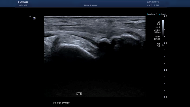
An ultrasound examination performed on a Canon Medical Aplio a550 system using an 18L7 high-frequency transducer demonstrated an unusual appearance of the tibialis posterior tendon. A large incidental os tibiale externum (os navicular) variant was identified with normal synchondrosis articulation with the navicular (Video 1).
A small amount of fluid in the tibialis posterior tendon sheath was visualised and assessed to be greater than normally expected physiologically. The fat planes around the tibialis posterior tendon sheath are echogenic, which suggests the presence of a regional inflammatory response (Video 2).
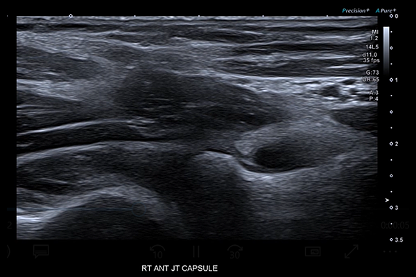
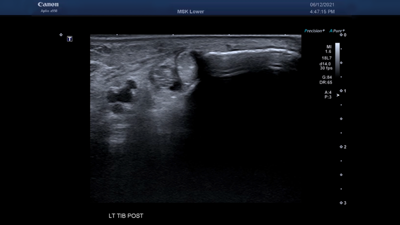
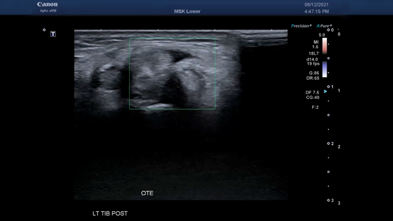
Within the tibialis posterior tendon, a very long and linear echogenic appearance was identified (Video 3). This appearance represents crystal deposition within the tendon. The high, low-flow sensitivity and excellent spatial and temporal resolution of the Advanced Dynamic Flow (ADF) technology on the Aplio a550 provided clear demonstration of hypervascularity within the tendon, the tendon sheath and adjacent fat planes. ADF also allowed clinicians to confirm the presence of a crystal deposition process by demonstrating some twinkle artifact arising from the echogenic material (Video 4, 5).
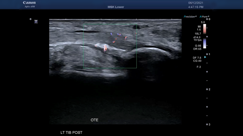
A faint line of crystal deposition corresponding to the sonographic findings was seen on dorsi-plantar foot and anterior-posterior ankle radiographs (Figure 1, 2).
The clinical presentation combined with sonographic and radiographic findings are characteristic of calcific tendinosis (HA deposition). Hydroxyapatite is a bone salt and requires an enthesis to migrate from bone to tendon. In this case, the enthesis is the large accessory os tibiale externum ossicle. The radiographic density of the crystal deposition suggests a diagnosis of hydroxyapatite over gout and the clinical presentation is consistent with an acute tendinosis event.
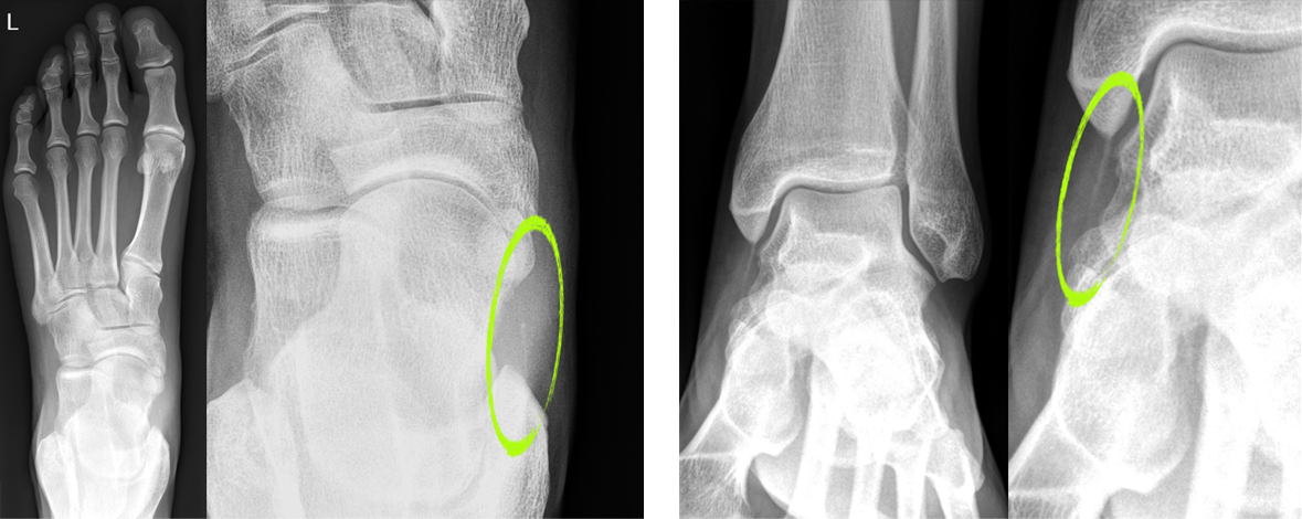 Figure 1, 2: DP foot and AP ankle radiographs demonstrate a faint line of crystal deposition, corresponding to the sonographic findings.
Discussion
Canon Medical’s Aplio a550 systems provide clinicians with high-quality B-mode imaging, which enables users to differentiate between similar density structures and better visualise abnormalities such as crystal deposition.
Advanced Dynamic Flow (ADF), available on all Aplio ultrasound systems, offers high-resolution colour flow that enables physicians to identify small complex vascularity. It offers superior spatial resolution to easily reveal minute blood flow patterns and provides high frame rates while maintaining the full B-mode image quality to accurately depict flow with directional information, even in tiny vessels.
The use of ADF in this clinical case clearly demonstrated hypervascularity within the tendon as well as confirming the presence of crystal deposition by twinkle artifact.
References
1. Basic calcium phosphate crystal-associated musculoskeletal syndromes: an update
Ann K. Rosenthal [Read here]
2. Detection of calcium phosphate crystals in the joint fluid of patients with osteoarthritis – analytical approaches and challenges
Alexander Yavorskyy,a,* Aaron Hernandez-Santana,a Geraldine McCarthy,b and Gillian McMahon [Read here]
Other Aplio a550 Case Studies by Stephen Bird
Social Media


