

Deep Learning Spectral
A fully integrated end-to-end spectral workflow
The Aquilion ONE / PRISM Edition harnesses the temporal benefits of rapid kV switching with patient specific mA modulation and combines them with a Deep Learning Reconstruction that delivers excellent energy separation and low-noise properties.
What’s more, its fully integrated end-to-end workflow is easy to use and can be conveniently incorporated into your routine protocols.
Whitepaper
Deep Learning Spectral CT
– Faster, easier and more intelligent
Kirsten Boedeker, PhD, DABR, Senior Manager, Medical Physics*1
Mariette Hayes, Global CT Education Specialist, Healthcare IT*1
Jian Zhou, Senior Principal Scientist*2
Ruoqiao Zhang, Scientist*2
Zhou Yu, Manager, CT Physics and Reconstruction*2
- 1 Canon Medical Systems Corporation
- 2 Canon Medical Research USA
Whitepaper
Deep Learning Spectral CT – Faster, easier and more intelligent
Kirsten Boedeker, PhD, DABR, Senior Manager, Medical Physics*1
Mariette Hayes, Global CT Education Specialist, Healthcare IT*1
Jian Zhou, Senior Principal Scientist*2
Ruoqiao Zhang, Scientist*2
Zhou Yu, Manager, CT Physics and Reconstruction*2
- Canon Medical Systems Corporation
- Canon Medical Research USA
Case study
Deep Learning Spectral: See beyond the metal artifacts
Prof. Mickaël Ohana MD, PhD
Nouvel Hôpital Civil,
Strasbourg University Hospital, Strasbourg, France
White paper
Diving into the Future of CT with Deep Learning Spectral Imaging
Prof. Catherine Roy
Head of the Radiology department,
Strasbourg University Hospital, France
New spectral applications integrated with the Aquilion ONE / PRISM Edition
Images can be delivered directly to your reading station, and with our range of new Vitrea applications, you can easily analyse comprehensive spectral data, including quantification and multi-layered images for a more detailed and definitive diagnosis.
Smartly developed, these applications provide a seamless solution from scan to diagnosis, and are scalable from a single workstation to a hospital-wide network, including PACS integration.
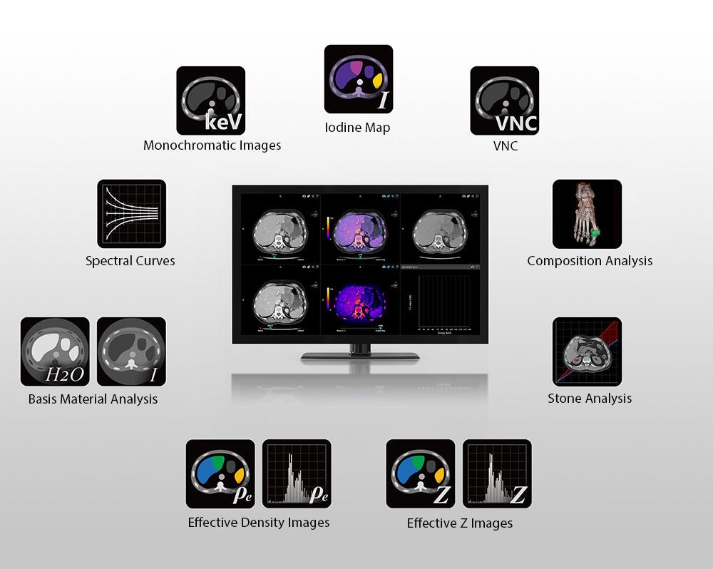
Vitrea Advanced Visualization*
Expand your spectrum with our latest healthcare IT solutions
Review Spectral data quickly and easily with a range of applications that simplify your workflow and provide clinically relevant features and outputs for every patient.
- Create effective monochromatic images for 166 energy levels ranging from 35 keV to 200 keV.
- View quantitative colour-coded iodine distribution maps that show the iodine uptake in anatomical structures.
- Characterise and differentiate tissue with effective Z and Electron density information at your fingertips.
- Differentiate between uric acid and non-uric acid urinary stones to make better-informed treatment decisions.
- Visualise and quantify the presence of monosodium urate in anatomical structures.
- Differentiate iodine from an intracranial haemorrhage.
- Vitrea is available as an option.
The content reflects information available at the time of publication and may differ from current information.
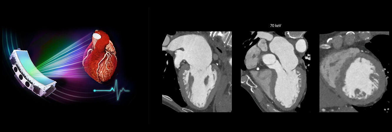
ECG gated volumetric Deep Learning Spectral cardiac CT
Technological Advantages
- 16 cm volumetric coverage
- One beat cardiac acquisition
- SEMAR
- Temporal congruency
- Spatial alignment
Clinical advantages
- Low keV to improve iodine conspicuity and reduce iodine contrast
Social Media


