Search

Image
Menu

Get the full picture when it matters most
With advanced ultrasound technologies such as iDMS, SMI, TSO and MicroPure, sharp and clear breast images with incredible detail provide a more confidence diagnosis.

Image

Intelligent Dynamic Micro-Slice

iBeam provides superior image quality whilst maintaining high frames rates

iDMS sharpens the ultrasound beams in the direction of the transducer lens. iDMS transducer elements are controlled separately to create a thin slice beam with continuous focus from near field to far field.

Image

The new iBeam architecture consists of three technologies that work together to cumulatively optimise the efficiency of the beam.

  • Multi-Sync Pulser for transmission of multiple signals
  • Multi-Beam Receiver enables the reception of these signals
  • Multi-Harmonic Processor providing unprecedented beam processing power
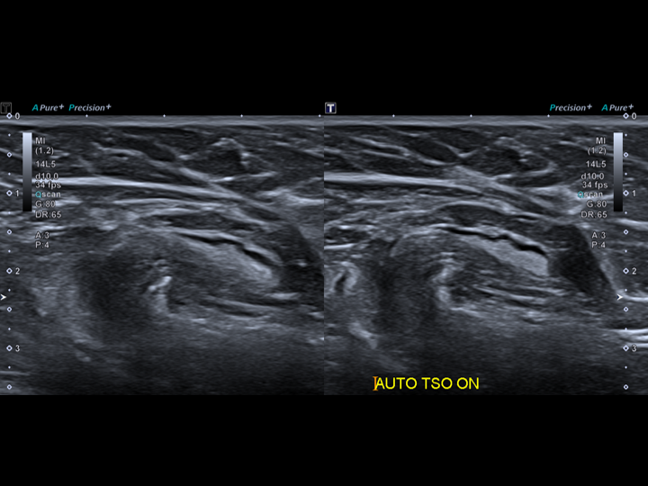
Automatic Tissue Specific Optimisation (TSO)

Automatically optimises with the touch of a button to compensate for variations in speed and sound across patient tissue types – providing focussed and high-resolution images.

Image

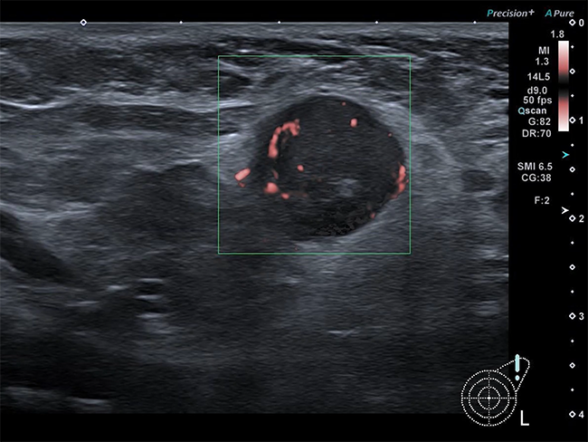
Superb Micro Vascular Imaging (SMI)


Diagnose breast lesions with unsurpassed confidence. SMI is a doppler technique designed to improve the visualisation of blood flow. The new adaptive algorithm removes clutter while maintaining a high frame rate which increases blood flow detection and depicts the microvascular architecture of breast lesions.

Image

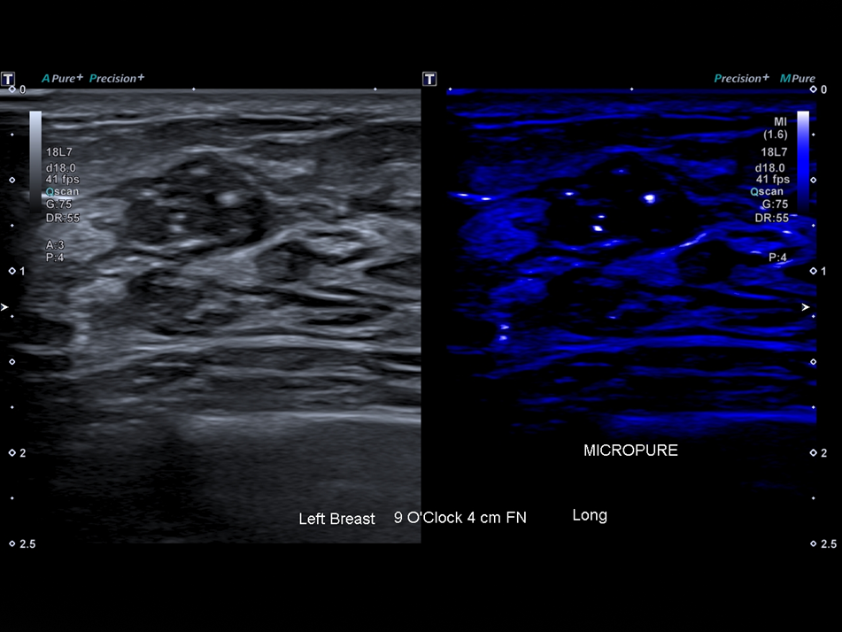
MicroPureTM

Automatically highlights microcalcifications (a potential marker for malignancy) in the breast and other organs.

Image

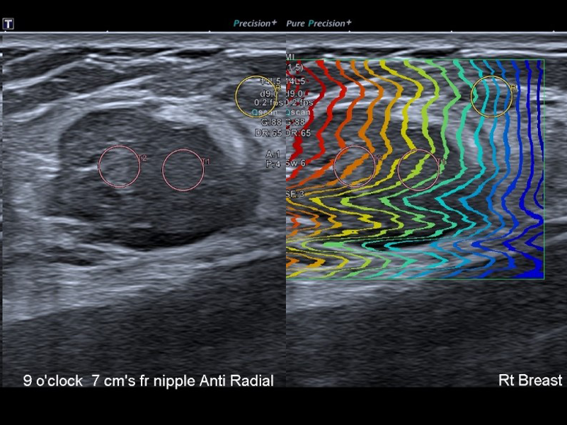
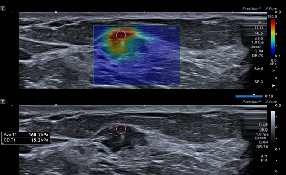
iPerformance – Shear Wave Elastography

Localise and assess palpable masses with high accuracy, sensitivity and reproducibility with Canon’s comprehensive Elastography solution. Identify the full extent of the lesion for accurate surgical planning.

Image

Advanced Dynamic FlowTM – ADF

Visualise micro-vasculature and complex flow patterns with unprecedented accuracy and detail.

Image

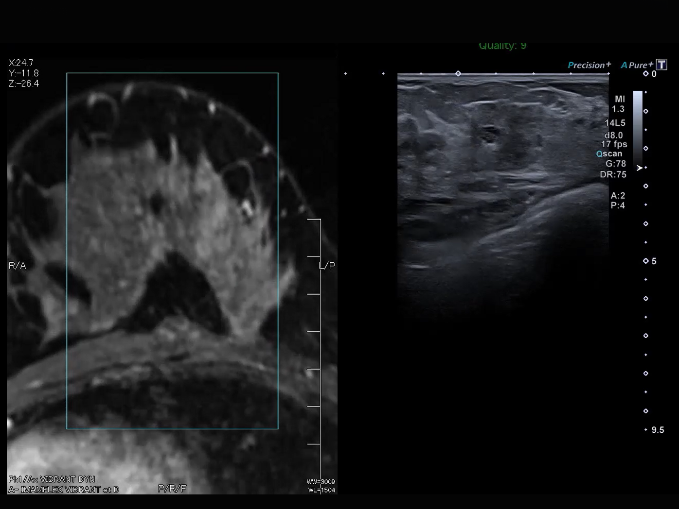
MRI-Ultrasound Fusion

Quick and easy multi-modality fusion to provide increased accuracy and confidence in ultrasound-guided procedures.


Image

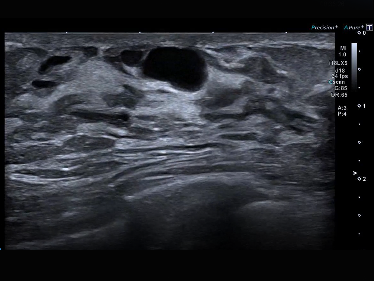
Precision Imaging

Precision Imaging reveals more clinical detail with significantly sharpened outline of lesions, enhanced image uniformity and reduced noise.

Image

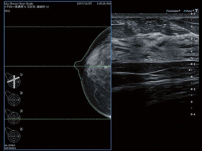
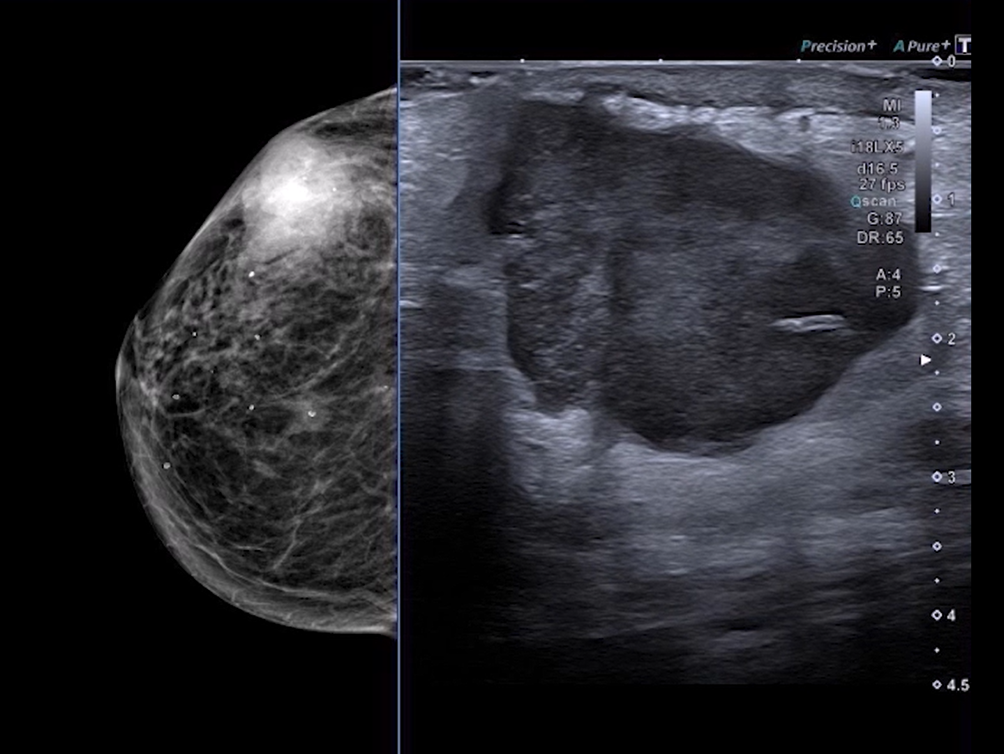
Breast Scan Guide

Combine real-time ultrasound imaging with mammography data for improved localisation of lesions.

Image

Differential Harmonics

See detail at all depths with Canon’s unique Differential Harmonics technology. Combining the pulse subtraction tissue harmonic frequency and the fundamental frequency for improved clinical confidence.

Image

Reference Mode

Combine real-time ultrasound imaging with pre-acquired mammography data within a region of interest for improved lesion localisation.

Image

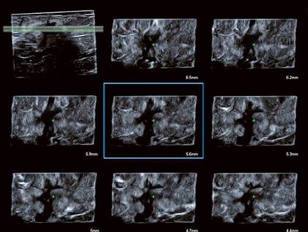
iPerformance – Smart 3D

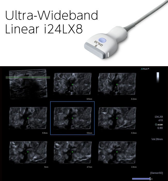
Achieve a more comprehensive analysis of lesion characteristics with the ability to easily reconstruct multiplanar images through a breast lesion.

Image

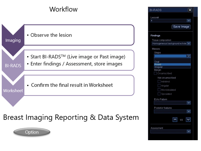
BI-RADSTM

Standardised structured reporting to increase efficiency.

Image

Breast Imaging

Standard and specialised ultra-wideband transducers with intelligent Dynamic Micro-Slice technology provide outstanding homogeneity and superior definition across the entire field of view.

Image

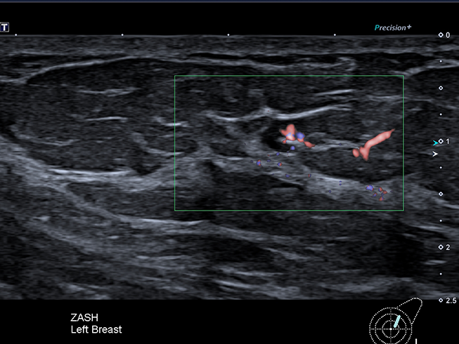
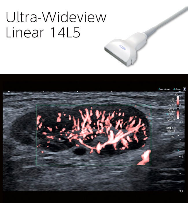
Advanced technologies like SMI are available on specialty transducers for advanced exams such as the quantitative assessment of inflammation in lymph nodes.

Image

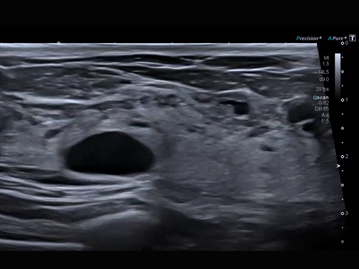
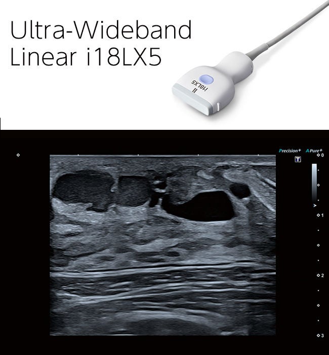
The outstanding resolution of this high frequency transducer helps identify fine details and differentiate between anatomical structures with ease.

Image

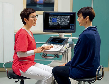
Consistent superior image quality across a range of clinical applications makes Canon’s Women’s Health range valuable for comprehensive examinations.

Image