

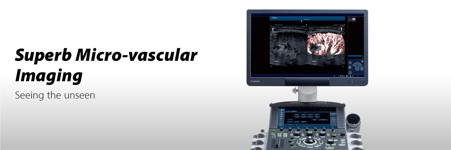
Superb Micro-vascular Imaging (SMI)
Seeing the unseen
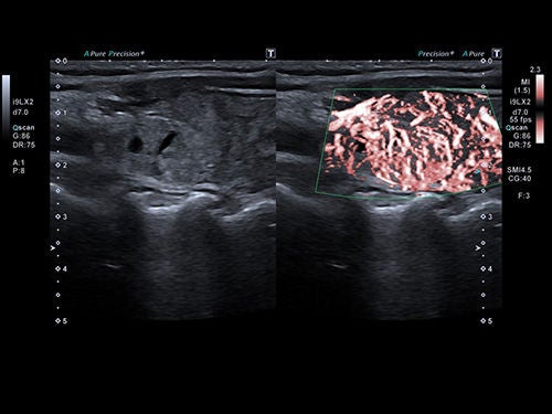
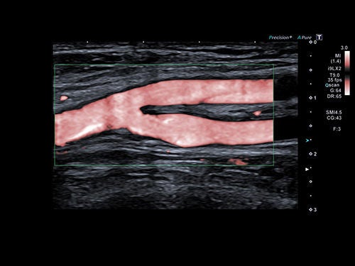
SMI is a technology that expands the range of visible blood flow and provides visualization of low microvascular flow never seen before with ultrasound. By enabling detailed visualization of vascular flow signals, SMI supports users during their clinical decision-making processes, reducing routine use of invasive methods.
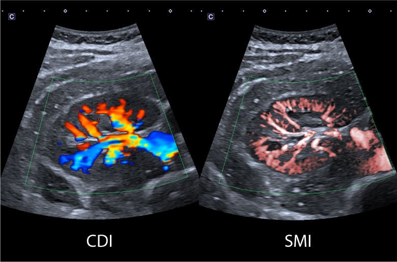
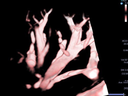
Three modes of SMI
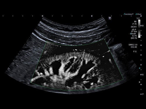
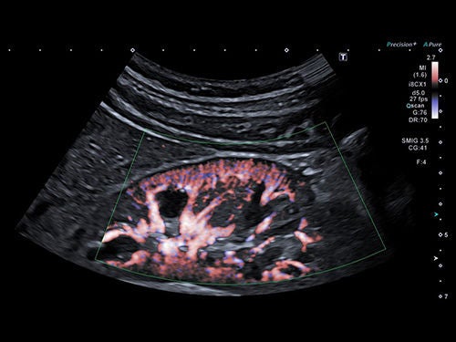
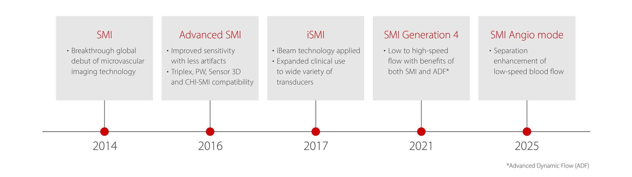
Evolution of SMI
SMI is an innovative ultrasound technology introduced in 2014.
In 2017, “iSMI” was introduced, allowing simultaneous processing of multiple signals to maintain both noise reduction and high frame rates, even when the ROI is expanded. In 2021, the technology evolved into “SMI Generation 4,” which extended the flow velocity range from low to high speed flows. This advancement enabled high velocity motion artifacts to be reduced and expanded the application of SMI to include the visualization of high velocity blood flow.
In 2025, SMI achieved further progress in visualizing low-velocity blood flow, marking a new stage in its development and expanding its clinical potential.
SMI Angio mode
Seeing the unseen – in finer detail
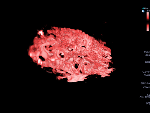
Building on SMI, Aplio’s new SMI Angio mode delivers superior blood flow visualization, offering finer detail and enhanced separation of flow in small, closely adjacent vessels, supporting more accurate assessment and increased diagnostic confidence in a wide range of use cases.
Clinical Images
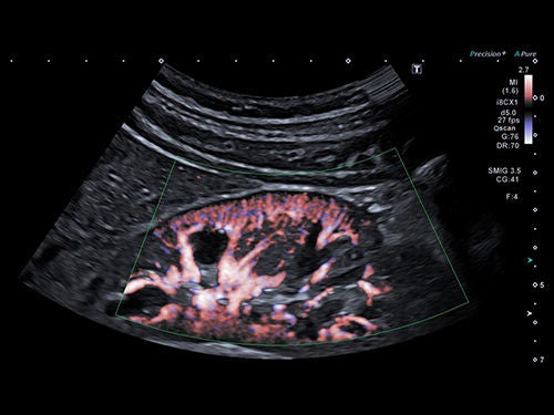
SMI proves to be useful in many clinical applications and anatomical regions.
SMI
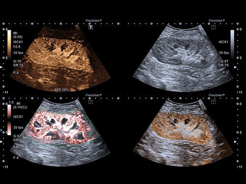
SMI Angio mode

Education and Resources



White Paper
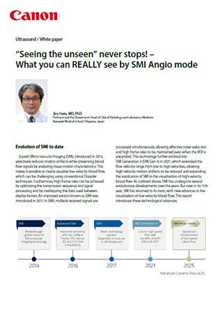
Prof. Jiro Hata
"Seeing the unseen" Never stops!- What you can REALLY see by SMI Angio mode
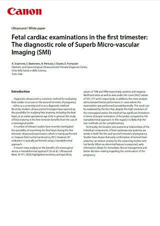
A. Sciarrone, S. Bastonero, A. Pertusio, I. Dusini, E. Fornaciari
Fetal cardiac examinations in the first trimester: The diagnostic role of Superb Micro-vascular Imaging (SMI)
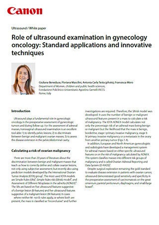
Giuliana Beneduce, Floriana Mascilini, Antonia Carla Testa, Francesca Moro
Role of ultrasound examination in gynecology oncology: Standard applications and innovative techniques
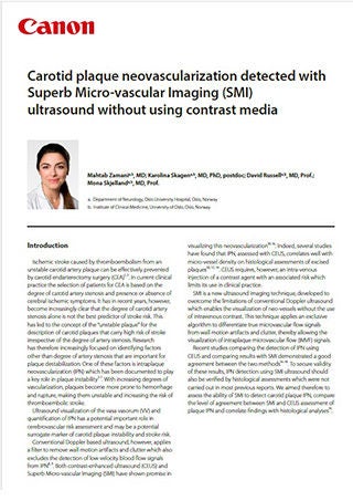
Mahtab Zamani, MD; Karolina Skagen, MD, PhD, postdoc; David Russell, MD, Prof.; Mona Skjelland, MD, Prof.
Carotid plaque neovascularization detected with Superb Micro-vascular Imaging (SMI) ultrasound without using contrast media
Interview report and Case study
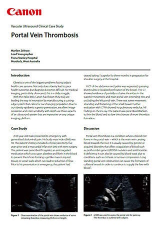
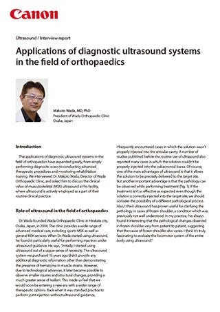
Makoto Wada, MD, PhD
Applications of diagnostic ultrasound systems in the field of orthopaedics
Akihiro Narita, MD, PhD
Norifumi Sawamukai, MD, PhD
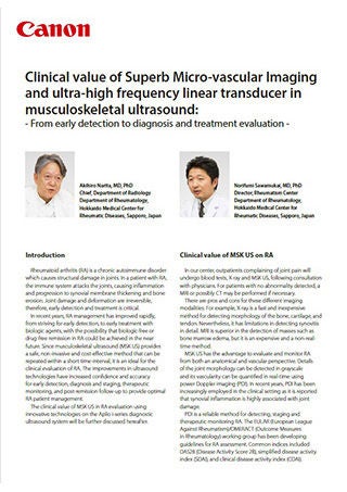
Clinical value of Superb Micro-vascular Imaging and ultra-high frequency linear transducer in musculoskeletal ultrasound:
Seeking more information? We can help.

Social Media


