

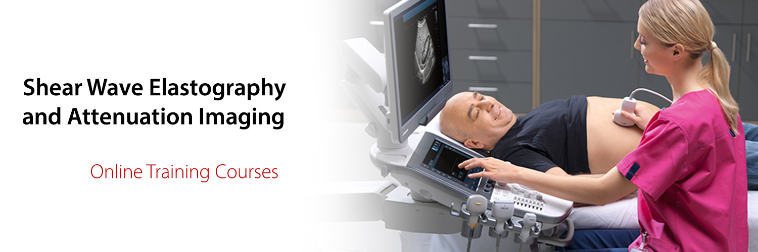
These courses have been developed to give ultrasound users an intensive primer in using quantitative chronic liver disease diagnostic functions.
This is a unique opportunity to learn with Marilyn Zelesco (Fiona Stanley Hospital) and Dr. Todd Erpelding.
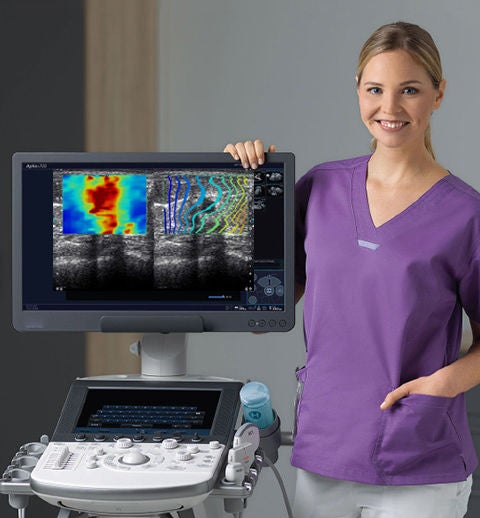
Shear Wave Elastography (SWE) course
This advanced SWE ultrasound course includes 15 comprehensive modules, designed as a comprehensive primer for ultrasound practitioners to utilise the SWE function to quantify liver stiffness for cirrhosis grading. Access discussion and practical examples of SWE physics, scanning techniques and interpretation of results.
| Module1 | Introduction to SWE |
| Module2 | Physics of SWE with Dr. Todd Erpelding |
| Module3 | Patient preparation, scanning technique and reporting |
| Module4 | SWE considerations. Discussion of variations and confounders |
| Module5 | Practical demonstration of scanning technique |
| Module6 | Live scanning. Pitfalls and confounders |
| Module7 | Live scanning. Left lobe of liver pitfall |
| Module8 | Live scanning. Patient forced expiration pitfall |
| Module9 | Live scanning. How to achieve a stable elastogram |
| Module10 | Live scanning. Liver capsule confounders |
| Module11 | Live scanning. NAFLD patient demonstration |
| Module12 | Live scanning. Demonstration of NAFLD patient with geographical infiltration |
| Module13 | Case study. Alcoholic liver disease |
| Module14 | Case study. HC patient post treatment |
| Module15 | Conclusion |
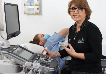
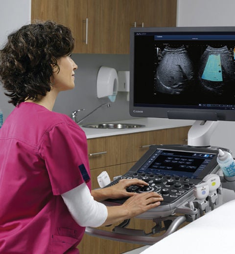
Attenuation Imaging (ATI) of the liver course
Nine comprehensive modules to help utilise ATI for liver steatosis quantification. Including discussion and practical examples of ATI physics, scanning technique, recognition of confounders and interpretation of results.
| Module1 | Reasons for attenuation imaging |
| Module2 | Reasons for attenuation imaging |
| Module3 | Patient preparation, technique, reporting and considerations |
| Module4 | Practical introduction to scanning techniques |
| Module5 | Practical demonstration of confounders in healthy patients |
| Module6 | Practical demonstration of NAFLD patient |
| Module7 | Practical demonstration of geographical fatty infiltrated liver |
| Module8 | Practical confounders in S2 geographical liver |
| Module9 | Practical demonstration of targeted ultrasound of the liver (TUSL) including SWE |

Social Media


